
On this page
Questions about your own assay?
Ask a specialistAntibody binding kinetics most often go wrong in the setup. A bivalent antibody in solution rebinds and makes the off-rate look slower than it is. Capture the antibody by its Fc, flow monomeric antigen over it at five concentrations around the expected KD, let dissociation run long enough, and report the setup with the numbers.
Antibody binding kinetics tell you more than KD
Antibody binding kinetics describe how fast an antibody finds its antigen and how long it stays bound. You measure them in real time on a label-free biosensor and fit the curves to get the rates.
The association rate constant (kon, in 1/M/s) says how fast the complex forms. The dissociation rate constant (koff, in 1/s) says how fast it falls apart. The equilibrium dissociation constant KD is koff divided by kon, and two antibodies with the same KD can have very different kinetics [1].
Make koff concrete. At a koff of 0.0001 1/s, half of the complexes are still intact after about 1.9 hours. The rates tell you how quickly an antibody engages its target and how long it holds on, which bears on its duration of action [1]. When you compare engineered antibody variants, the rates also show whether a mutation sped up binding or slowed release.
How do you measure antibody binding kinetics?
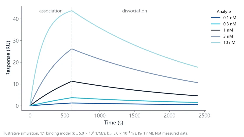
Immobilize one partner on a sensor surface, flow the other over it at several concentrations and record binding in real time. The rise during injection depends on both rates, the decay in buffer on koff alone. A global fit of a 1:1 model to the whole series gives kon, koff and KD [2].
Surface plasmon resonance (SPR), biolayer interferometry (BLI), grating-coupled interferometry (GCI) and Nanophotonic Evanescent field Sensing (NES) all work this way: each detects binding at the sensor surface without a label. Solution methods skip the surface. A kinetic exclusion assay measures free antibody in solution at equilibrium and is often used for sub-nanomolar affinities [1].
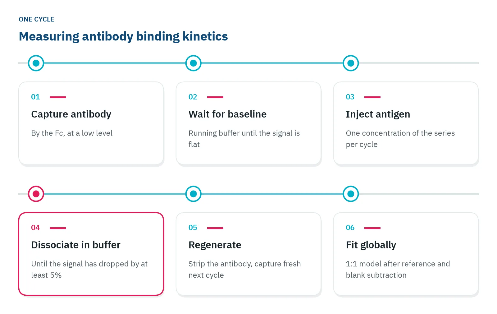
One measurement cycle on a label-free biosensor has six steps.
Capture the antibody, flow the antigen
An IgG has two binding arms. Flow it over a surface full of antigen and one arm can let go while the other still holds, so it rebinds. Dissociation looks slower and affinity looks higher than either arm really is [1,2].
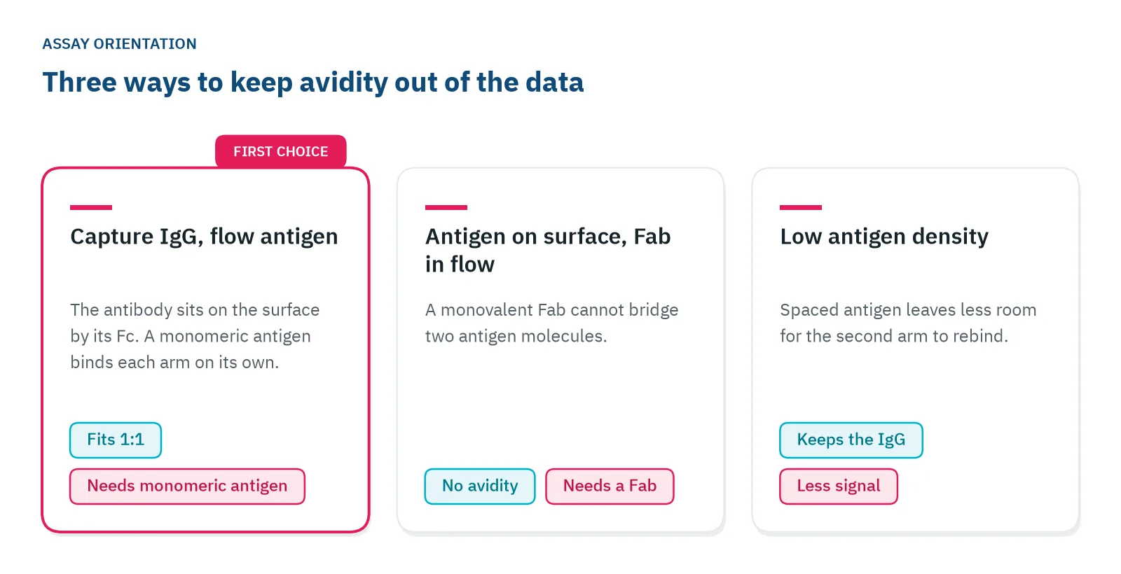
Reverse the orientation when you can. Capture the IgG and flow a monomeric antigen over it, so each arm binds on its own and a 1:1 model holds [2,3]. If the antigen must stay on the surface, flow a monovalent Fab instead [2], or lower the antigen density, which reduces bridging but also the signal [3].
Capture by the Fc keeps every antibody oriented the same way, with its binding arms free [4]. Protein A, Protein G and anti-Fc antibodies all capture this way, and Delta supplies sensor chips with Protein A and Protein G surfaces for this format.
Then check the capture itself. A capture reagent with weaker hold on the antibody may make the antigen off-rate look faster than it is [5]. Run one blank cycle: capture the antibody, inject buffer, watch the signal. What you lose there is capture decay. Subtract it from every cycle.
Regenerate Protein A or G surfaces with a short injection of 10 mM glycine at pH 1.5 to 2, and run kinetics at a flow rate around 30 µL/min. Protein A and G bind IgG subclasses and species with different strength, so test the capture with your own antibody.
Set concentrations and times from the expected rates
Inject the antigen as a dilution series from 0.1 to 10 times the expected KD. Run five or more concentrations and inject one of them twice as a reproducibility check [6]. For an antibody with a KD near 1 nM, that means about 0.1 to 10 nM.
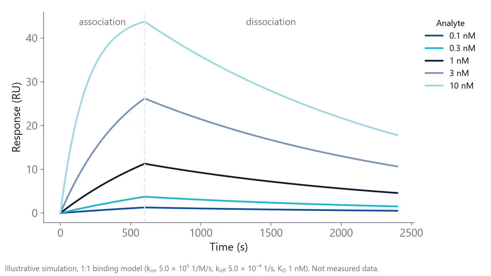
Then set the times. The association phase needs visible curvature. The dissociation phase must lose at least 5% of the signal, or koff cannot be measured accurately [4]. Work out what that means for your antibody: at a koff of 0.00001 1/s, a 5% decay takes about 85 minutes. In one comparison of high-affinity antibodies, the flow-based instruments ran 10 minutes of association and 45 minutes of dissociation [5]. That is enough for 5% decay only when koff is above about 2 × 10⁻⁵ 1/s.
Two formats run the series. Multi-cycle kinetics regenerates and recaptures between concentrations. Single-cycle kinetics injects rising concentrations with short breaks and one long dissociation at the end, so it needs only one regeneration per series [4]. In the same comparison, single-cycle kinetics on Fc-captured antibodies gave rate constants close to those of the other platforms [5].
Check the fit before you trust the rates
Fit all concentrations together with a 1:1 model, with kon, koff and Rmax as global parameters [2]. Then look at the residuals. They should scatter around zero without a pattern.
Mass transport is the classic trap in antibody binding kinetics on a dense surface: antigen binds faster than the flow can bring it in, and kon comes out too low.
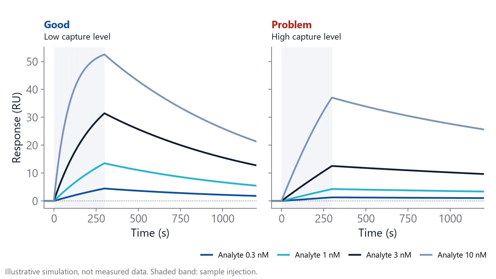
Repeat the series at a second, lower capture level. If kon and koff stay the same, transport is unlikely to limit them, though a density change can also alter the surface itself [4]. A higher flow rate is a weaker check on its own, because delivery grows only with the cube root of the flow rate [4].
If the fit stays poor, rule out the usual causes before you try a more complex model [6]:
- Aggregates in the antigen: spin or filter it before use
- Antigen sticking to the surface: check the signal on the reference channel
- Avidity: reverse the orientation or switch to a Fab
- Capture decay or drift: subtract a blank cycle
What should you report with antibody binding kinetics?
Report kon, koff and KD with the model you fitted, and enough of the setup for someone else to repeat it. Different instruments rank the same antibodies in much the same order but can differ in absolute values, so the setup is what makes your numbers comparable [7].
- Orientation: which partner is on the surface, the capture chemistry and the capture level
- Antigen concentrations, association and dissociation times, flow rate and temperature
- Number of replicates and the spread between them
- Fit model, which parameters were global and the residuals or chi-squared
- Any koff that decayed less than 5%, given as an upper limit, not a value
On inQuiQ
Antibody binding kinetics on inQuiQ
High-affinity antibodies need a long, stable dissociation phase before koff can be read at all. inQuiQ measures dissociation rate constants down to 10⁻⁶ 1/s, with baseline drift typically below 0.3 RU/min. In the standard setup, capture a different antibody on Protein A or Protein G in three channels and keep the fourth as the reference. One antigen injection flows through all four channels in series, so you get three antibodies against the same antigen sample, reference-subtracted, before the surface is regenerated. The tight binders application note shows antibody kinetics with a short and a long dissociation protocol.
Questions researchers ask
Is an EC50 from ELISA the same as KD?
An EC50 is the antibody concentration that gives half the maximum signal in that particular assay, not an affinity. It depends on antigen coating density, avidity, washing and incubation time, so it can sit far from the true KD. Use it to rank antibodies within one assay, and measure kinetics when you need the affinity itself.
Which concentration do I enter for an IgG in a solution assay?
With a monomeric antigen in solution, the two arms of an IgG behave as independent binding sites. Enter twice the molar IgG concentration as the concentration of binding sites when you fit. This correction does not apply to avidity on a dense antigen surface; there, change the orientation instead.
Can you rank antibody clones by off-rate without knowing their concentration?
Yes. koff does not depend on how much antibody is captured, so you can capture clones straight from culture supernatant, inject one antigen concentration and compare the dissociation curves. This gives a ranking, not a KD. For kon and KD you still need purified antibody of known concentration.
At what temperature should you measure antibody kinetics?
25 °C is a common standard, which makes results easy to compare between labs. Rates change with temperature, so a value at 25 °C is not the value at 37 °C. If you need behavior closer to the body, add a run at 37 °C and report the temperature with every number.
References
- Lodge J, Kajtar L, Duxbury R et al.. Quantifying antibody binding: techniques and therapeutic implications. MAbs. 2025. doi:10.1080/19420862.2025.2459795
- Real-Time Analysis of Biomolecular Interactions. Springer Japan. 2000. doi:10.1007/978-4-431-66970-8
- Apiyo DO. Biolayer Interferometry (Octet) for Label-free Biomolecular Interaction Sensing. Handbook of Surface Plasmon Resonance. 2017. doi:10.1039/9781788010283-00356
- Handbook of Surface Plasmon Resonance. The Royal Society of Chemistry. 2017. doi:10.1039/9781788010283
- Yang D, Singh A, Wu H et al.. Determination of High-affinity Antibody-antigen Binding Kinetics Using Four Biosensor Platforms. J Vis Exp. 2017. doi:10.3791/55659
- Marquart JA. SPRpages – Getting a Feeling for the Curves. Handbook of Surface Plasmon Resonance. 2017. doi:10.1039/9781788010283-00106
- Yang D, Singh A, Wu H et al.. Comparison of biosensor platforms in the evaluation of high affinity antibody-antigen binding kinetics. Anal Biochem. 2016. doi:10.1016/j.ab.2016.06.024
Keep reading
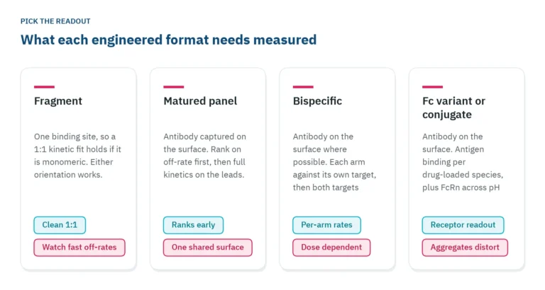 Antibody engineering: what binding data each format needsFragments, bispecifics, Fc variants and conjugates each need a different binding readout. Which parameter to measure, in which assay orientation, and why valency decides both.Applications
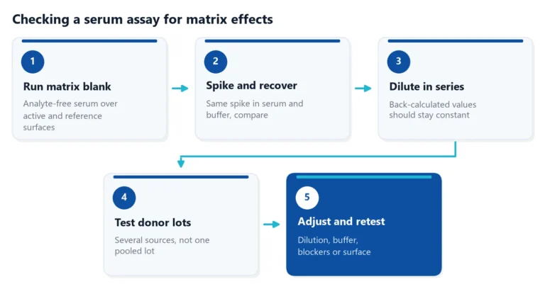 Matrix effects in binding assays: how serum changes your signalSerum can raise, lower or blur the signal of a binding assay. Learn where matrix effects come from, how to test for them, and how to reduce them in ELISA and label-free assays.Applications
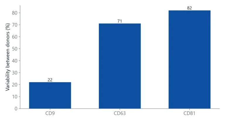 Tetraspanins CD9, CD63 and CD81: EV markers and binding targetsTetraspanins CD9, CD63 and CD81 are the standard handles for detecting and capturing extracellular vesicles. This article explains what they are, why they are not on every vesicle, and how to set up binding measurements around them.