
On this page
Questions about your own assay?
Ask a specialistTetraspanins are four-pass membrane proteins, and CD9, CD63 and CD81 are the most used markers for extracellular vesicles (EVs). They are not on every vesicle, and their mix differs by source and donor. The tetraspanin you capture with decides which EVs you measure, so profile your source first and choose capture targets with care.
What are tetraspanins?
Tetraspanins are a family of small membrane proteins with four transmembrane domains, a short and a long extracellular loop, and 33 members in mammals. They group other membrane proteins into clusters and guide their transport. CD9, CD63 and CD81 are the members most researchers meet, because they are standard markers of extracellular vesicles (EVs) [1].
The protein backbone is 200 to 300 amino acids long, with variable glycosylation. Cysteines inside the cell carry palmitoyl groups, and cysteines in the large extracellular loop (LEL, also called EC2) hold its fold together. In crystal structures of CD9 and CD81, the four helices form a cone with a cavity inside the membrane, and the LEL rests on top. In CD81 this cavity held a cholesterol molecule. Other structures show an open form, in which the LEL reaches away from the membrane [1].
For binding work, the LEL is the part to know. It is exposed outside the membrane and it is flexible: CD81 crystal structures show slightly different LEL shapes, which may help CD81 bind partners such as the hepatitis C virus E2 protein. Tetraspanins also bind each other and proteins such as integrins, CD19 and ADAM10, forming the so-called tetraspanin web. Many of these contacts are indirect, so co-precipitation alone does not prove a direct interaction [1].
Why CD9, CD63 and CD81 mark extracellular vesicles
Tetraspanins are among the most abundant membrane proteins on EVs [2]. That is why antibodies against CD9, CD63 and CD81 are a standard way to detect, capture and classify vesicles. The three differ in where they sit in the cell. CD63 is found mostly in late endosomes and multivesicular bodies, so it tends to mark exosomes, which form there. CD9 and CD81 sit mostly at the plasma membrane and decorate ectosomes, which bud from it. The split is not strict: CD63 also appears on ectosomes [1].
The difference is useful. Small EVs contain exosomal and non-exosomal subpopulations, and immuno-isolation with CD63, CD81 or CD9 pulls out different ones. Classic markers such as flotillin and HSP70 are found on all EV types alike [3].
What tetraspanins do inside EVs is less clear. Studies of EV release, cargo sorting and uptake after loss of CD9, CD63 or CD81 give conflicting results. A 2025 review concludes that these three do not play a general role in sorting cargo into EVs and are not essential for EV formation [1]. For assay design, treat them as handles on the vesicle, not as proof of a mechanism.
Are tetraspanins found on every extracellular vesicle?
Tetraspanins are on many EVs, but not on all of them. CD9, CD63 and CD81 are spread unevenly across single EVs. Each source has its own pattern of which tetraspanins appear together. That pattern stays the same across isolation methods but differs between sources. Some vesicles carry one tetraspanin, some carry several, and some may carry none that capture antibodies recognize [4].
Size plays a part too. In EVs from one cell line, particles of 100 nm or smaller were more often positive for CD9 and CD81 than larger ones. Platforms that only count tetraspanin-captured EVs therefore see a skewed size distribution [5].
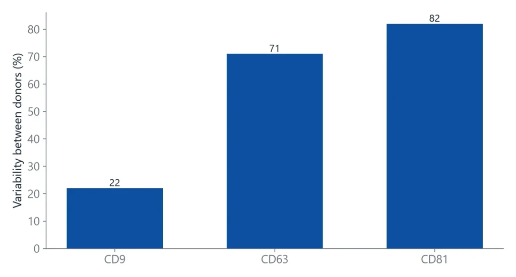
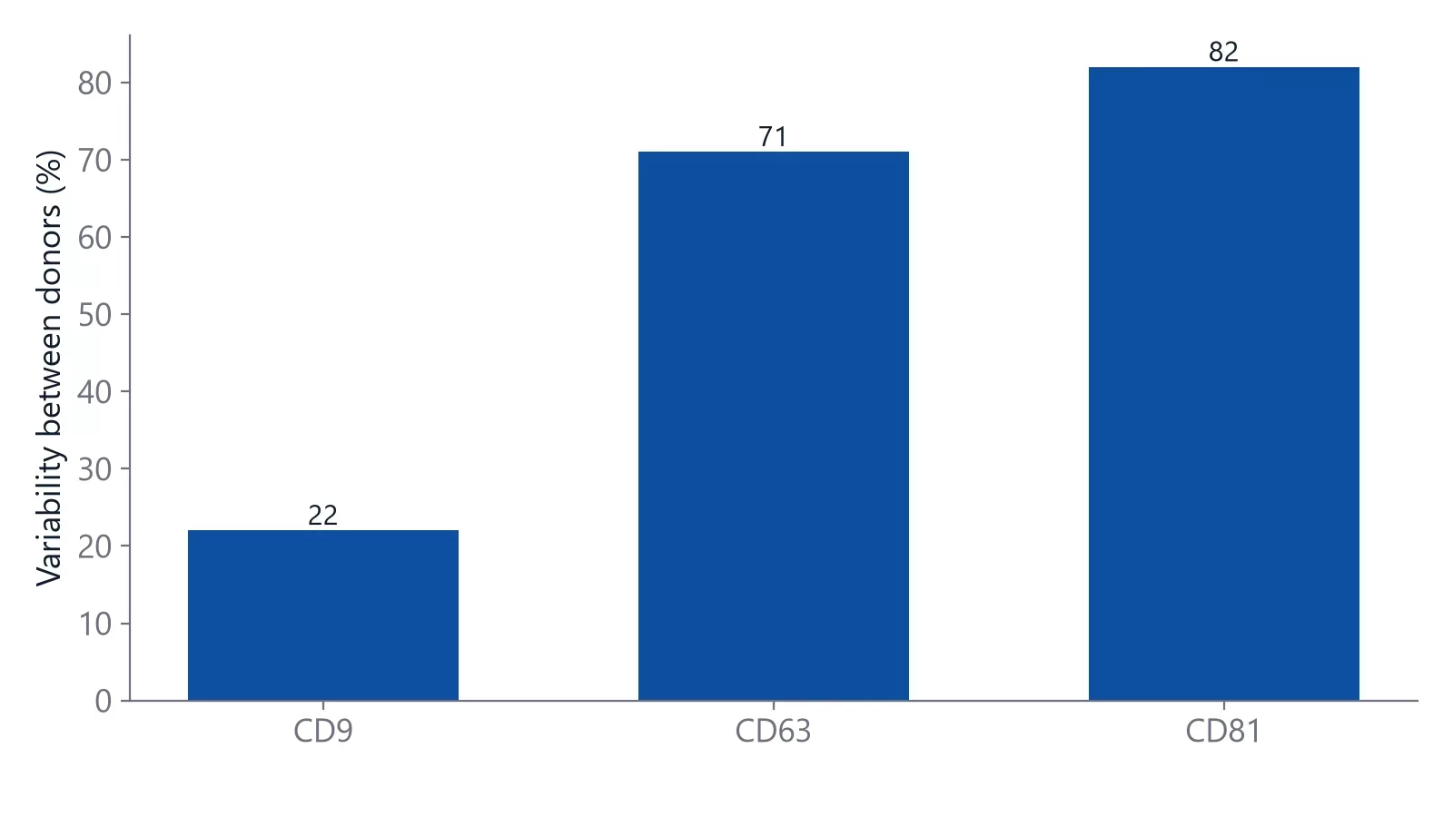
Profiles also change between cell types and between people. A bead-based assay found clear differences across 13 cell lines: EVs from U118 glioblastoma cells carried mostly CD9, while U87 EVs carried more CD81 and CD63. In plasma from healthy donors, CD9 made up a median of about 72% of the total tetraspanin signal, and CD63 and CD81 levels varied much more between donors than CD9 [6].
Plasma and serum also add matrix effects, so plan reference surfaces as described in binding analysis in serum and plasma.
How your capture choice shapes what you measure
Many EV assays start with a tetraspanin capture step, in ELISA, bead-based flow cytometry and chip formats [4]. That step is a filter. In a study of ovarian cancer cell EVs, CD24-positive vesicles appeared only after CD9 capture or after non-specific capture of biotinylated EVs. Non-specific capture found about ten times more EpCAM-positive EVs and about five times more HER2-positive EVs than the tetraspanin capture spots [4].
How large the bias is depends on the source. In the same study, serum EVs were captured as well by anti-CD9 as by non-specific capture. EVs from placental mesenchymal stem cells were different: non-specific capture bound more of them than any single tetraspanin did, and their profile matched none of the single-tetraspanin surfaces [4].
Combining capture antibodies helps. On a surface plasmon resonance (SPR) sensor carrying two anti-tetraspanin antibodies on one surface, EV detection was higher than predicted from single-antibody surfaces. Tuning the ratio of the two antibodies improved sensitivity further. The authors link this to tetraspanin-enriched microdomains, where several tetraspanins sit close together [7].
The practical rule follows from both studies. Before you pick one tetraspanin as capture target, profile your EV source with all three, and report which one you used. On a 4-channel label-free biosensor such as inQuiQ, all three can be read in one injection. Tetraspanin-independent capture gave a broader view of the population [4], and a surface with two anti-tetraspanin antibodies detected more EVs than single-antibody surfaces [7].
How do you measure binding to tetraspanins on EVs?
There are two formats. To get a KD, immobilize a recombinant tetraspanin or its LEL and flow the antibody or partner protein over it as the analyte. To study vesicles, immobilize an anti-tetraspanin antibody and flow EVs over it. The second format gives relative binding or EV concentration, not a clean KD.
The first format is an ordinary protein interaction. It follows the usual rules of label-free binding analysis: a reference surface, a concentration series and a fit to a binding model, as in any antibody affinity and kinetics study. Because the LEL is flexible, check that the recombinant loop is folded, and confirm key results with a second construct.
The second format behaves differently. An EV is a particle of tens to hundreds of nanometers with many copies of each tetraspanin, so one vesicle can bind several capture antibodies at once. Multivalent binding, mass transport and a mix of binding sites all push curves away from the 1:1 model, and rebinding makes the off-rate look slower than it is [8]. A simple test is to compare two capture densities. If the curves change shape with density, transport, rebinding or uneven binding sites are likely part of what you see [8].
A way around this is to measure concentration instead of affinity. One group captured exosomes on an anti-CD63 SPR surface and used the diffusion-limited binding rate to estimate EV concentration. They had to correct for the broad size distribution and for vesicles about as large as the roughly 150 nm SPR probing depth, and reached an accuracy better than ±50% [9].
Setting up a tetraspanin capture experiment
A capture assay for EVs works best when each step answers one question. The workflow below keeps capture bias and non-specific signal visible.
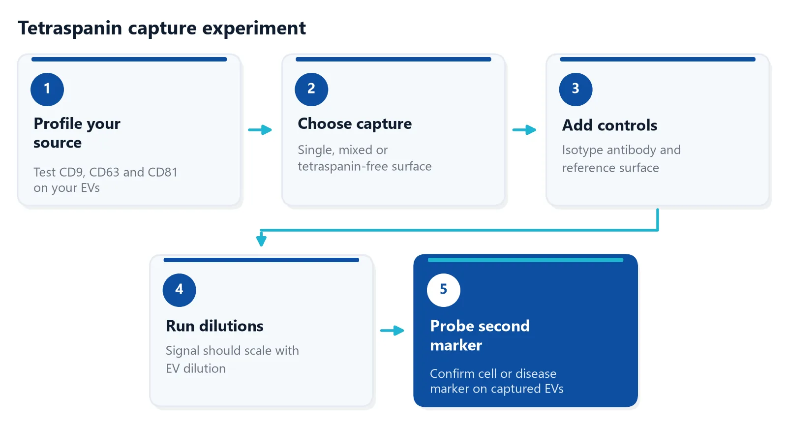
Two details are easy to miss. First, EV preparations contain non-vesicle particles, so describe how you separated and characterized them; the MISEV2023 guidelines list the options and their limits [10]. Second, check that signal scales with dilution. In one single-EV capture study, counts no longer doubled when the EV concentration doubled above about 5 × 10^8 particles/mL, so single spots may have held more than one vesicle [4].
Tetraspanin capture panels on inQuiQ
On the inQuiQ benchtop biosensor, each of the 4 channels carries its own capture surface: anti-CD9, anti-CD63 and anti-CD81 antibodies, with an isotype control as the reference channel. Each EV sample flows through all four channels in series, so one injection shows how the vesicles bind each tetraspanin and the control before the surface is regenerated.
The sensor surface is antifouling, which supports measurements in complex matrices such as serum and plasma [11]. Our LNP and EV characterization page describes how this applies to vesicle and nanoparticle work.
Questions researchers ask
Does biotinylating EVs change their tetraspanin profile?
In a single-EV study by Mizenko and colleagues, biotinylating surface amines and capturing EVs on anti-biotin hardly changed the apparent tetraspanin profile in most cases. The catch is that biofluids contain free proteins and lipoproteins that also take up biotin and then compete for the capture surface. Separate EVs from non-vesicle particles before you biotinylate.
Why do flow cytometry and capture assays give different tetraspanin profiles?
They see different vesicles. Scatter-triggered flow cytometry detects only larger EVs, from about 100 nm in the setup Mizenko and colleagues calibrated, while capture assays only see EVs that carry the capture protein. Fluorophores and unbound antibody also differ. Treat each profile as specific to its method, and report the method with the result.
Can a CD9 antibody change EV behavior in a functional assay?
It can. As summarized in a 2025 review by Rubinstein, pre-incubating fibroblast-derived EVs with a CD9 antibody reduced their uptake by pancreatic cancer cells, but it is unclear whether CD9 itself caused the effect. Other quantitative work found that loss of CD9 or CD63 did not change delivery of EV contents. Always include an isotype control antibody in blocking experiments.
Are tetraspanins only found on extracellular vesicles?
According to a 2025 review by Rubinstein, tetraspanins also sit on cell membranes, membrane protrusions, migrasomes and midbody remnants, and CD9 and CD81 have been found on HIV and influenza virus particles. In samples that may contain enveloped viruses, a CD9 or CD81 capture surface can bind more than EVs. Use a second marker to confirm what you captured.
References
- Rubinstein E, Théry C, Zimmermann P. Tetraspanins affect membrane structures and the trafficking of molecular partners: what impact on extracellular vesicles?. Biochem Soc Trans. 2025. doi:10.1042/BST20240523
- Andreu Z, Yáñez-Mó M. Tetraspanins in extracellular vesicle formation and function. Front Immunol. 2014. doi:10.3389/fimmu.2014.00442
- Kowal J, Arras G, Colombo M et al.. Proteomic comparison defines novel markers to characterize heterogeneous populations of extracellular vesicle subtypes. Proc Natl Acad Sci U S A. 2016. doi:10.1073/pnas.1521230113
- Mizenko RR, Brostoff T, Rojalin T et al.. Tetraspanins are unevenly distributed across single extracellular vesicles and bias sensitivity to multiplexed cancer biomarkers. J Nanobiotechnology. 2021. doi:10.1186/s12951-021-00987-1
- Fagúndez P, Olivera Á, Gololobova O et al.. Tetraspanin Positivity as a Function of Extracellular Vesicle Size Measured by a Modified Immuno-TEM Protocol. ACS Appl Bio Mater. 2025. doi:10.1021/acsabm.5c00591
- Tordoff E, Allen J, Elgart K et al.. A novel multiplexed immunoassay for surface-exposed proteins in plasma extracellular vesicles. J Extracell Vesicles. 2024. doi:10.1002/jev2.70007
- Lopez Baltazar JM, Gu W, Yu Q. Enhancing Extracellular Vesicle Detection via Cotargeting Tetraspanin Biomarkers. Anal Chem. 2024. doi:10.1021/acs.analchem.4c04086
- Handbook of Surface Plasmon Resonance. The Royal Society of Chemistry. 2017. doi:10.1039/9781788010283
- Rupert DL, Lässer C, Eldh M et al.. Determination of exosome concentration in solution using surface plasmon resonance spectroscopy. Anal Chem. 2014. doi:10.1021/ac500931f
- Welsh JA, Goberdhan DCI, O'Driscoll L et al.. Minimal information for studies of extracellular vesicles (MISEV2023): From basic to advanced approaches. J Extracell Vesicles. 2024. doi:10.1002/jev2.12404
- Delta Life Science. Biomarker Detection in Complex Clinical Matrix. Application note. Link
Keep reading
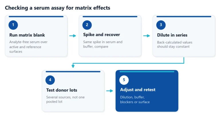 Matrix effects in binding assays: how serum changes your signalSerum can raise, lower or blur the signal of a binding assay. Learn where matrix effects come from, how to test for them, and how to reduce them in ELISA and label-free assays.Applications
 How to measure nanobody binding affinity and kineticsNanobodies often bind in the nanomolar to picomolar range. This guide covers which method to use, how to avoid avidity and off-rate traps, and how to check that a nanobody KD is reliable.Assay setup
 Baseline drift in a biosensor: why it happens and how to stop itBaseline drift is often blamed on the chip. A short buffer-only test shows whether the surface, the buffer or the instrument is the real cause.