
On this page
Questions about your own assay?
Ask a specialistELISA reads one endpoint after several washes, so it shows what stayed bound, not how fast molecules bind and let go. Binders that dissociate quickly can wash away unseen. When you need KD and rate constants, the alternative to ELISA is a real-time, label-free biosensor; for affinity alone, a solution method works. Keep ELISA for concentrations.
ELISA measures what survives the washes
An enzyme-linked immunosorbent assay (ELISA) coats one partner on a plate, adds the other, washes, and turns the bound amount into a color with an enzyme-labeled antibody. It is cheap, sensitive and runs in almost every lab.
As a binding study, it has one blind spot: you read a single number at the end. Every wash, and every incubation without free analyte, gives complexes time to fall apart. Candidates that dissociate fast or bind weakly can be lost in the washes, and the signal cannot tell association from dissociation [1].
A titration curve does not fix this. An EC50 from a plate depends on coating density, antibody valency and timing, so it ranks binders under your conditions but is not a KD. In one comparison, solid-phase assays made the antibodies look 1.5 to 5.4 times tighter binders than solution measurements, and the factor changed from antibody to antibody [2].
Two binders with the same KD can behave very differently
The dissociation constant KD tells you how tightly a complex holds at equilibrium, in molar units: lower is tighter. It is the ratio of two rates, koff divided by kon. The association rate constant kon (1/M/s) says how fast the complex forms. The dissociation rate constant koff (1/s) says how fast it falls apart.
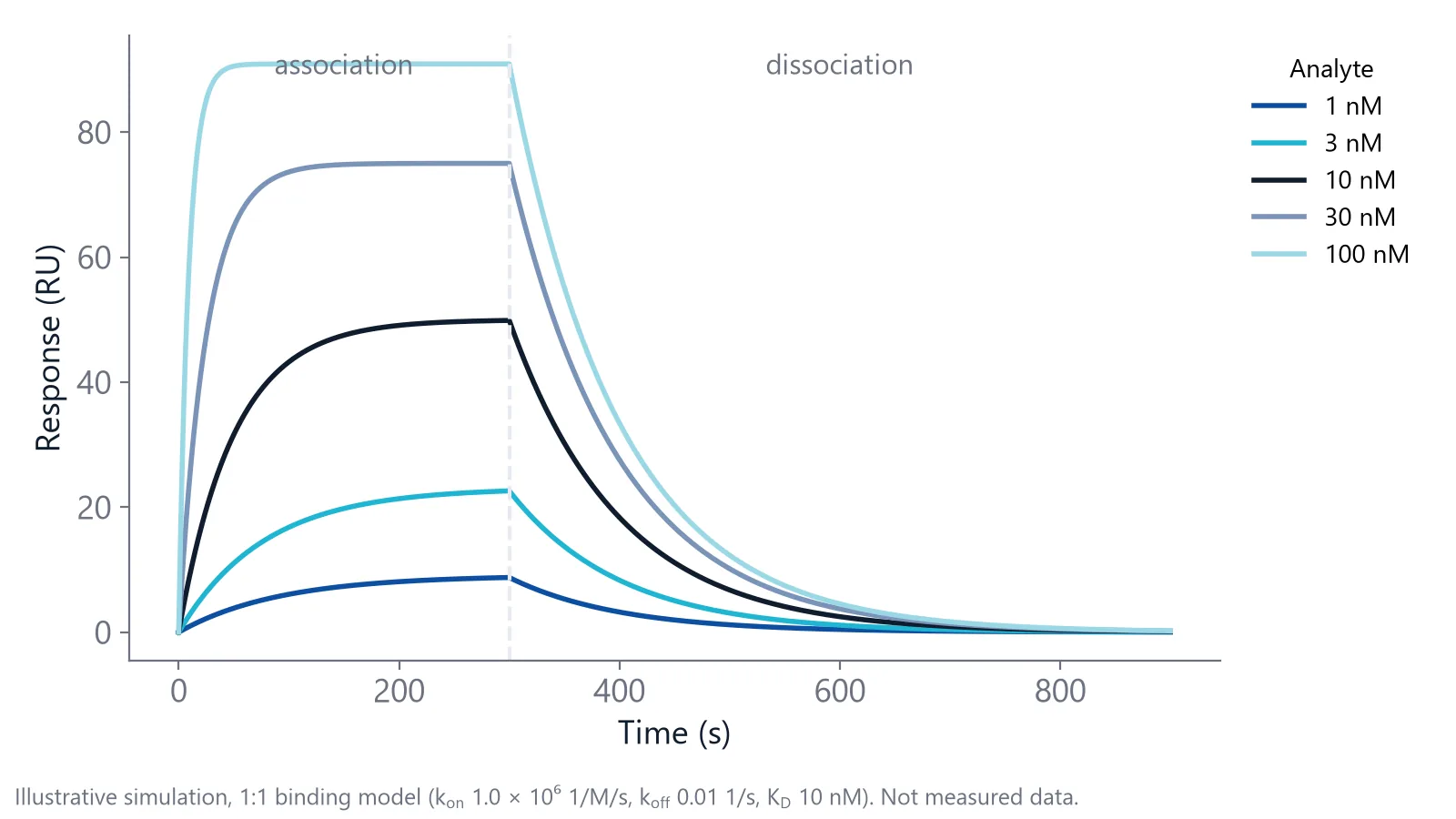
Take two antibodies, both with a KD of 10 nM. The first has a kon of 106 1/M/s and a koff of 0.01 1/s. Half of its complexes are gone about 70 seconds after the analyte is removed, and after five minutes of washing about 95% are gone.
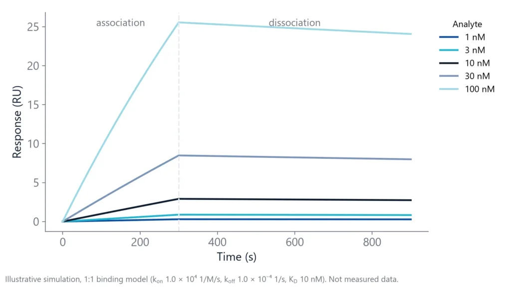
The second has a kon of 104 1/M/s and a koff of 10-4 1/s. Its complexes have a half-life of almost two hours, so five minutes of washing removes about 3%.
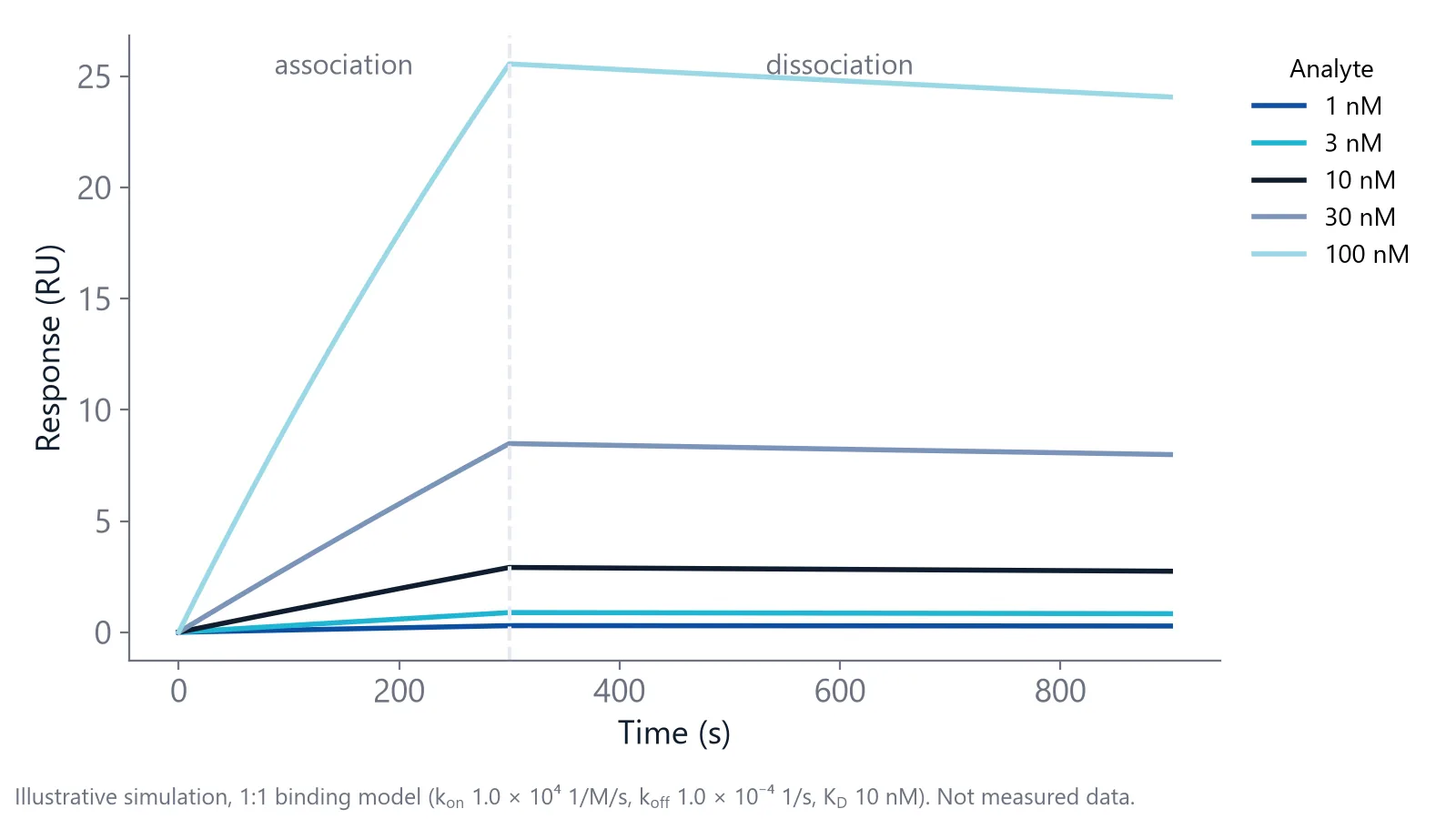
On a plate, the first antibody looks much weaker than the second. In a drug program the difference can matter more than the KD: a long residence time on the target can mean a longer effect and better selectivity [3]. For how this plays out in antibody selection, see antibody affinity and kinetics.
Which alternative to ELISA fits your binding question?
If you need rates, use a label-free biosensor, which watches binding at a surface as it happens. For affinity in solution, isothermal titration calorimetry (ITC) and microscale thermophoresis (MST) are the usual choices [5]. To stay on plates, a solution-equilibrium ELISA gives a true KD, but no rates [6].
Label-free biosensors include surface plasmon resonance (SPR), biolayer interferometry (BLI), grating-coupled interferometry (GCI) and Nanophotonic Evanescent field Sensing (NES). One partner, the ligand, sits on the sensor. The other, the analyte, is brought over it in solution, and the signal rises as mass binds and falls as it leaves. NES and SPR share one principle: binding changes the refractive index, how strongly the material slows light, in a thin field at the sensor surface. Read more on how photonic biosensing works.
| Method | What you get | Label | Watch out for |
|---|---|---|---|
| Label-free biosensors (SPR, BLI, GCI, NES) | kon, koff and KD from one series; also concentration | None; one partner is immobilized | Immobilization and regeneration need development |
| ITC | KD plus enthalpy and entropy, in solution [4] | None | No rate constants in a standard run |
| MST | KD in solution, low protein use, no immobilization [4] | One partner fluorescent | The label can cause artifacts [4] |
| Solution-equilibrium ELISA | KD in solution, even with crude antibody [6] | Enzyme-labeled detection antibody | No rates; antigen must be in large excess [6] |
| Standard ELISA | Yes or no binding, concentration, relative ranking | Enzyme-labeled detection antibody | Endpoint only; fast-dissociating binders can be lost [1] |
An SPR assay can take longer to establish than a simple ITC run [4]. NES and the other biosensors need the same surface development. The methods also check each other: confirming a KD with a second technique gives you more confidence in it [4].
A real-time readout finds binders that ELISA washes away
The format changes which binders you detect, as well as how much data you get. A clinical study tested patients for antibodies against the therapeutic antibody panitumumab with two assays: a bridging ELISA and a real-time biosensor assay [7].
On paper, the ELISA was 100 times more sensitive: it detected 10 ng/mL of a control antibody, against 1 µg/mL for the biosensor. Yet it found developing antibody responses in 2 of 612 subjects, while the biosensor found 25 of 604, including 8 with neutralizing antibodies [7].
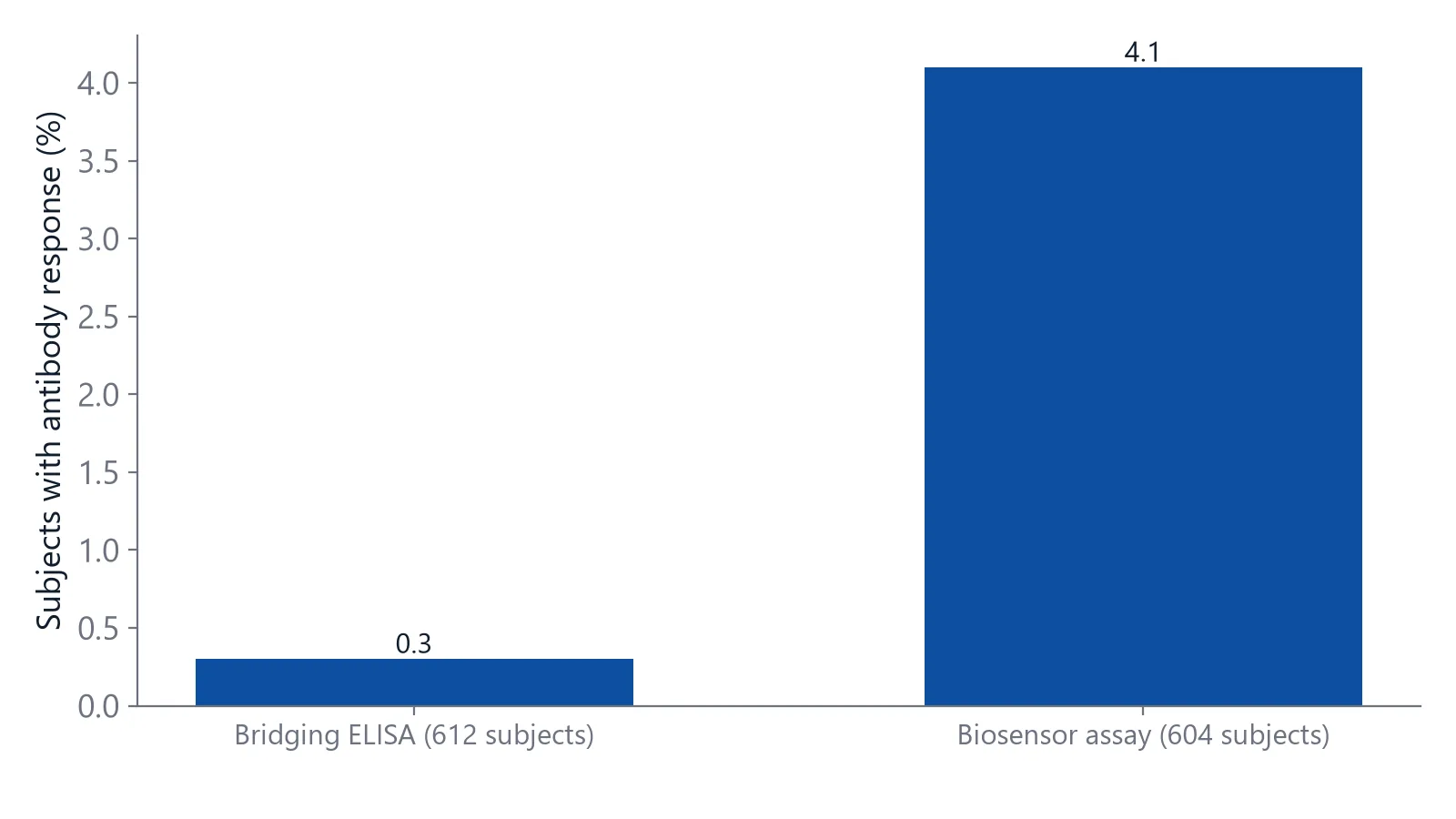
Tests with control antibodies of known affinity point to a reason: the ELISA was more sensitive for high-affinity antibodies, the biosensor for low-affinity ones [7]. The two formats see different parts of the affinity range. The study used SPR; BLI, GCI and NES also read binding while the analyte is still present, with no wash before the read.
If an ELISA negative matters for a decision and low-affinity binders are plausible, check it with a real-time method before you rely on it.
How your ELISA steps translate to a label-free run
Moving to a label-free alternative to ELISA keeps most of what you know. The coating protein usually becomes the ligand, your sample becomes the analyte, and the detection step disappears because the signal is the binding itself.
| ELISA step | Label-free step | What changes |
|---|---|---|
| Coat the plate | Immobilize or capture the ligand on the sensor | Use a capture tag where you can, so the binding site stays free |
| Block | Block the surface and keep a reference surface without ligand | The reference reads buffer effects and non-specific binding |
| Incubate the sample | Inject a concentration series of analyte | Association is recorded live |
| Wash | Switch back to buffer | Dissociation is recorded and gives koff |
| Detection antibody and substrate | None | No secondary reagents |
| Standard curve | Fit a binding model to all curves together | Gives kon, koff and KD |
Surface preparation happens once; the last three phases repeat in every cycle.
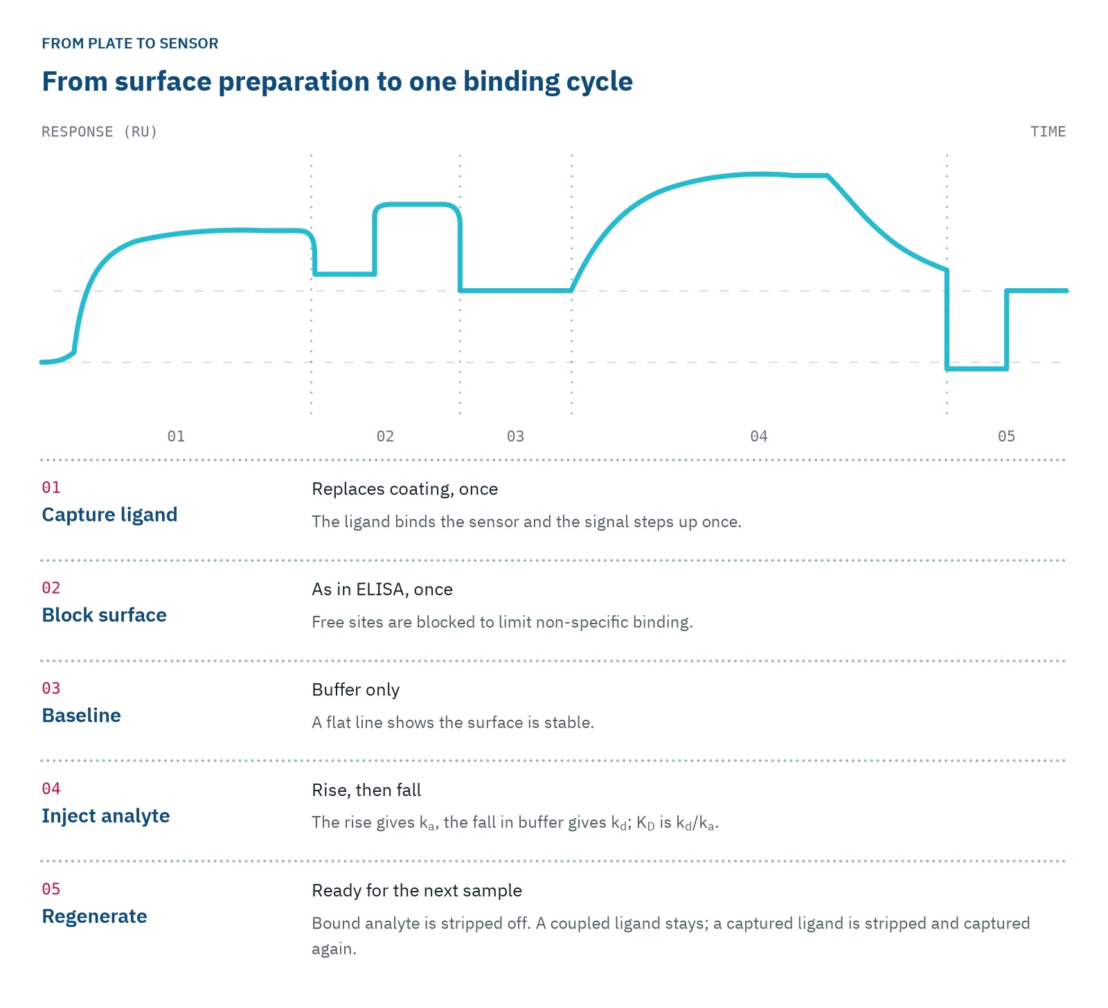
Before your first full run:
- Inject five analyte concentrations from about 0.1 to 10 times the KD you expect
- Let dissociation run until the signal has clearly dropped; a slow binder needs far longer than a fast one
- Repeat identical injections with regeneration in between, and check that the response and the baseline return to the same level [1]
On the inQuiQ biosensor, one injection flows through all 4 channels in series, one of them the reference, so a single sample reads up to 3 ligands plus a reference.
When is ELISA still the right tool?
ELISA is still the right tool when you need to know how much analyte a sample holds and binding rates will not change your decision. Before you look for an alternative to ELISA, check whether your case is one of these:
- Concentrations in hundreds of samples with a validated kit
- Very low levels of a high-affinity analyte, where enzyme amplification wins (as in the panitumumab study)
- Labs with a plate reader and no access to a biosensor
Biosensors can measure concentration too. Like an ELISA, they capture the analyte from solution, but they read it from the binding signal itself, with no detection step [8]. An inQuiQ application note shows IL-6 analysis in under 3 minutes [9].
A common split works well: screen on plates, then measure kinetics on the shortlist. Send forward only the clones that pass, as purified protein, so the first kinetic fit is clean.
Questions researchers ask
Does a sandwich ELISA tell you anything about affinity?
Only indirectly. The signal depends on two antibodies holding the analyte through several washes, so both affinities and both off-rates shape it. A strong signal shows that the pair works at your concentrations. It cannot give the KD of either antibody; measure each one separately for that.
Can I use my ELISA reagents on a biosensor?
The binders transfer; some of the buffer does not. A blocking protein or carrier protein in a kit buffer can stick to a label-free surface and add signal, and a sample buffer that differs from the running buffer causes a bulk shift. Exchange samples into the running buffer and check kit buffers for additives before you inject.
Why does my biosensor ranking not match my ELISA ranking?
Start with the checks. Sort your candidates by koff and compare that order with the plate. Then test the antibody as a monovalent Fab, or at low ligand density, to see whether avidity explains the gap. Concentration and incubation time on the plate also shift a ranking. If the koff order explains the mismatch, both data sets are probably right.
Do I need purified protein for a label-free assay?
For the ligand, often you do not. Capture through a tag or a specific antibody pulls it out of a mixture, and the reference surface subtracts much of the background. For the analyte, purity matters if you want exact rates, because aggregates and other binders add signal. Use purified material for your first kinetic fit.
References
- Real-Time Analysis of Biomolecular Interactions. Springer Japan. 2000. doi:10.1007/978-4-431-66970-8
- Azimzadeh A, Pellequer JL, Van Regenmortel MH. Operational aspects of antibody affinity constants measured by liquid-phase and solid-phase assays. J Mol Recognit. 1992. doi:10.1002/jmr.300050103
- Copeland RA, Pompliano DL, Meek TD. Drug-target residence time and its implications for lead optimization. Nat Rev Drug Discov. 2006. doi:10.1038/nrd2082
- Holdgate G, Embrey K, Milbradt A et al.. Biophysical methods in early drug discovery. ADMET DMPK. 2019. doi:10.5599/admet.733
- Walport LJ, Low JKK, Matthews JM et al.. The characterization of protein interactions - what, how and how much?. Chem Soc Rev. 2021. doi:10.1039/d1cs00548k
- Friguet B, Chaffotte AF, Djavadi-Ohaniance L et al.. Measurements of the true affinity constant in solution of antigen-antibody complexes by enzyme-linked immunosorbent assay. J Immunol Methods. 1985. doi:10.1016/0022-1759(85)90044-4
- Lofgren JA, Dhandapani S, Pennucci JJ et al.. Comparing ELISA and surface plasmon resonance for assessing clinical immunogenicity of panitumumab. J Immunol. 2007. doi:10.4049/jimmunol.178.11.7467
- Apiyo DO. Biolayer Interferometry (Octet) for Label-free Biomolecular Interaction Sensing. Handbook of Surface Plasmon Resonance. 2017. doi:10.1039/9781788010283-00356
- Delta Life Science. High-Throughput Analysis. Application note. Link
Keep reading
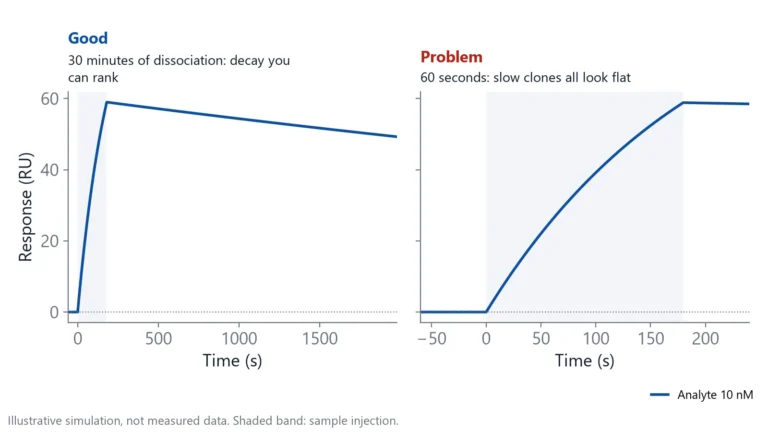 Antibody clone screening: from ELISA hits to off-rate rankingAn ELISA tells you which clones bind, not which ones hold on. How to rank hybridoma and library clones by off-rate straight from supernatant, and when ELISA is still the right first step.Switching from ELISA
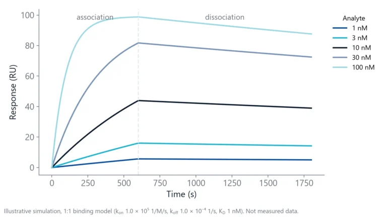 ELISA binding affinity: what it can tell you and what it missesELISA can estimate a KD, but the format decides what that number means. Four formats, three biases, the checks to run and when ELISA is still the right tool.Method comparisons
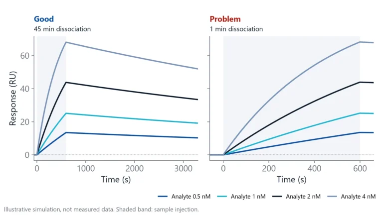 Biolayer interferometry vs SPR: which method fits your assayBoth methods measure binding label-free and in real time. The sample path, flow over a chip or a tip in a well, decides which one suits your hardest measurement.