
On this page
Questions about your own assay?
Ask a specialistELISA tells you which clones bind, but its washed endpoint signal mixes affinity with antibody concentration and avidity, so it cannot rank clones by how long they stay bound. Capture each antibody from supernatant on a label-free biosensor, inject the antigen once, and rank by off-rate. Keep ELISA for the first yes or no.
An ELISA hit says the clone binds, not how long it holds on
Antibody clone screening means testing the supernatants of many hybridoma or library clones to find the few worth keeping. Most labs start with an ELISA: antigen coated in a well, supernatant added, unbound material washed away, a labelled secondary antibody read as color.
That color says the clone binds. It does not say how long the antibody stays on the antigen. ELISA reads one endpoint after several washes, so it cannot separate association from dissociation, and clones that let go fast can be lost in the wash [1]. Three more things shape the signal:
- Antibody concentration, which is usually unknown in supernatant and cannot be adjusted for each clone [2]
- Avidity, because a bivalent IgG on a coated surface can look stronger than its single binding site [1,2]
- The coating itself, because antibodies raised against a hapten may recognize the linker of the coated conjugate rather than the free hapten [2]
A bright well can be a mediocre clone at high titer. A pale well can be an excellent clone that secretes little.
Why rank clones by off-rate?
Because the off-rate does not depend on how much antibody a clone secretes. In a clone screen, the amount a clone secretes changes only how much is captured, which sets the signal height. The rate at which a bound complex falls apart does not depend on concentration [1], which makes koff the most dependable number a single injection gives. kon and KD also come out after normalization but are less firm.
The off-rate, koff, is the fraction of complexes that dissociate per second (unit 1/s). Lower means a tighter hold. A koff of 10-3 1/s means about 0.1% of complexes fall apart each second, a half-life near 12 minutes. At 10-5 1/s the half-life is about 19 hours [1].
For a reagent or diagnostic antibody, the strongest binder often performs best, while a therapeutic candidate needs a balance with other properties [2]. What comes after the screen is covered on our page on antibody affinity and kinetics.
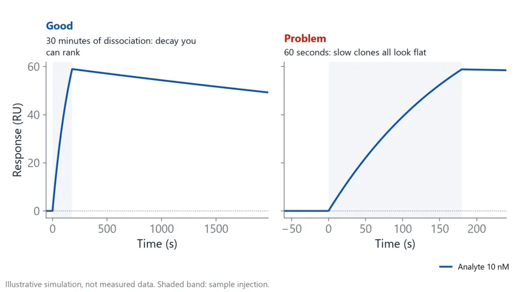
The dissociation phase must be long enough for the clones you hope to find. If almost every complex is still bound when you stop, the slow clones all look the same, and the best ones can be missed [1].
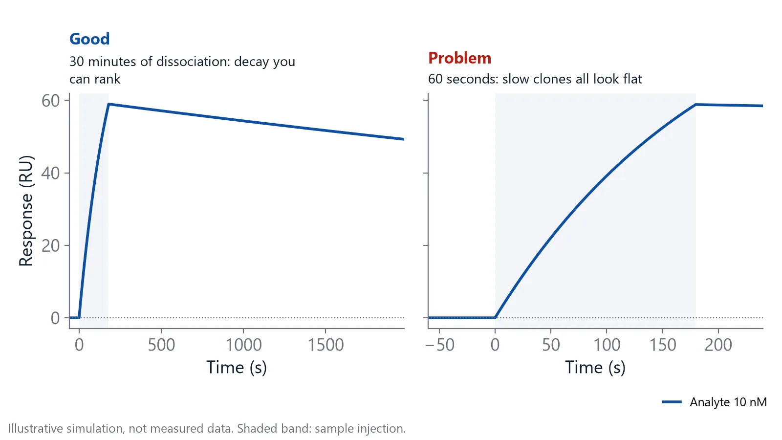
Pick the dissociation time from the slowest koff you want to resolve. At 10-4 1/s, 30 minutes of buffer removes about 16% of the complexes. At 10-5 1/s, it removes less than 2%.
Capture the antibody, inject the antigen
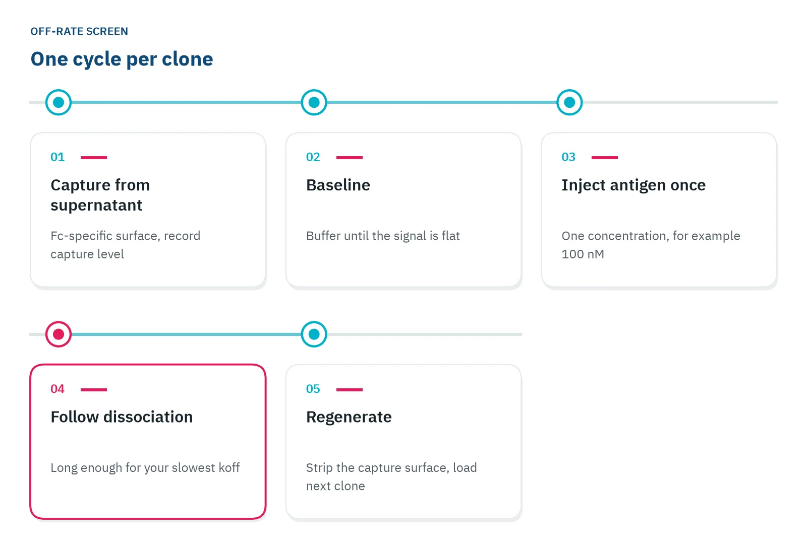
Label-free antibody clone screening usually turns the ELISA around. An Fc-specific surface captures each antibody from crude supernatant in the same orientation, and the antigen flows over in solution at a single concentration [3,4]. The surface does the purification for you.
When the antigen is monomeric, an antibody on the surface and antigen in solution give single-site binding [5], without the avidity that can inflate ELISA signals [1,2]. Dimeric or Fc-fusion antigens can still bind both arms of a captured IgG, so check the curve shapes as described below. Normalize each antigen response to the amount of antibody captured, then fit all clones with one model. You get kon, koff and KD for every clone under identical conditions [3]. Keep capture levels similar across clones so the comparison stays fair [6].
In one sensor comparison, a single-concentration kinetic assay used 100 nM antigen with 300 s association and 300 s dissociation, and the capture sensors were regenerated with 10 mM glycine at pH 1.7 [7]. Treat these as a starting point: 300 s resolves only fast clones (at 10-4 1/s about 3% of complexes dissociate), so follow the dissociation rule above. Filter the supernatant, dilute it in running buffer, and use spent medium diluted the same way for blank cycles.
Use a capture surface that holds on longer than your antibody does
The capture surface can distort the ranking. In one comparison, the same antibody gave a KD of 165 nM on one capture surface and between about 0.7 and 2 nM on the others, while a solution-based method gave a far tighter value still. Protein A can also bind the variable region of VH3 antibodies, not only the Fc [7].
If the antibody holds the antigen more tightly than the surface holds the antibody, the decay you record is the antibody leaving the surface. On a Protein A surface, the decay recorded was mainly that [7].
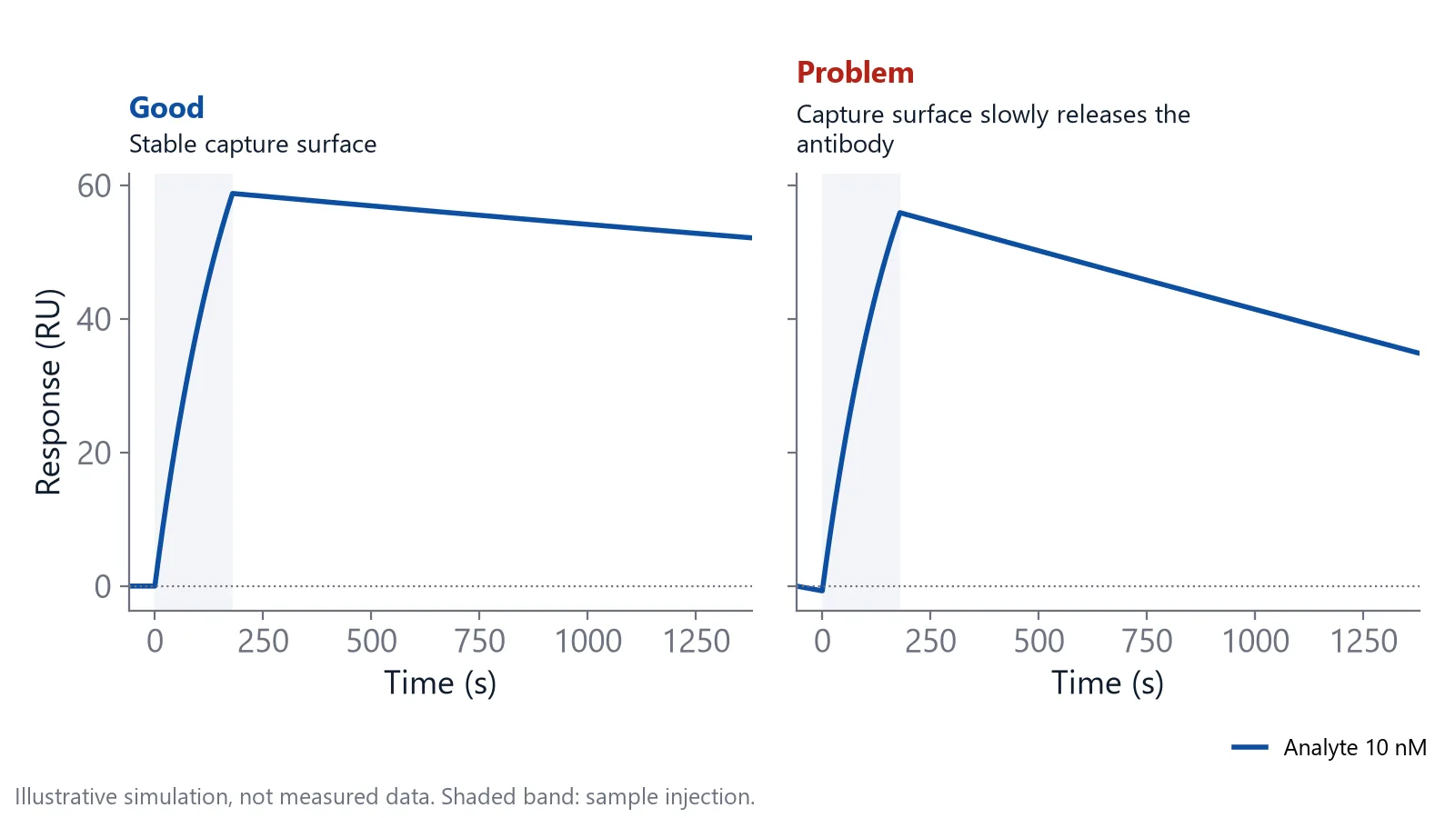
Run one blank cycle per capture surface: capture a known antibody, inject buffer instead of antigen, and watch the signal. What you lose there is capture decay, not dissociation. If it is as large as your slowest clone's dissociation, change the surface before you screen. Delta supplies sensor chips with Protein A and Protein G surfaces for Fc capture.
A screen ranks clones; it does not measure final constants
A single-concentration screen compares many antibodies in one run. Its numbers are good for ranking, not as definitive constants [6]. In the original protocol, single-concentration values agreed with follow-up runs at several antigen concentrations [3]. Repeat that check on your own top hits, at several antigen concentrations.
Before you trust the list, check:
- Capture level per clone, a rough reading of how much antibody the supernatant held (see antibody titer and quantification)
- A known reference antibody on every plate
- Curve shapes that point to bivalent binding, which a kinetic screen can flag [4]
Throughput is no longer the barrier it was. Published antibody clone screening protocols handle about 200 supernatants a day [3] up to several hundred antibodies a day [7], and off-rates can be read from crude bacterial lysates of Fab fragments [8].
When is ELISA still the right tool for antibody clone screening?
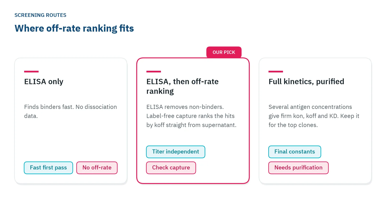
ELISA is still the right tool for the first yes or no across thousands of wells, when you have no biosensor, or when the final antibody will work in a coated-plate assay and you want to test it in that format. Add an off-rate ranking once the hit list is short enough that each choice matters.
Surface plasmon resonance (SPR), biolayer interferometry (BLI), grating-coupled interferometry (GCI) and Nanophotonic Evanescent field Sensing (NES) all read binding as it happens, without a label. NES is the photonic method behind Delta's instruments, and how photonic biosensing works explains the principle.
| ELISA | Label-free biosensors | |
|---|---|---|
| What you measure | One endpoint signal after washing | Binding and dissociation in real time: koff, and often kon and KD [3] |
| Label | Enzyme-labelled secondary antibody | None |
| Sample | Crude supernatant | Crude supernatant, antibody captured on the surface [3,7] |
| Throughput | Many plates a day on a plate reader | About 200 to several hundred samples a day in published protocols [3,7] |
| Strengths | Fast, low cost, standard lab equipment | Ranks by off-rate regardless of titer [1], single-site binding with monomeric antigen [5] |
| Limits | Mixes titer, avidity and wash loss [1,2] | Capture surface can distort data [7], ranks rather than gives final constants [6] |
Most teams end up with a two-step route.
On inQuiQ
Clone screening on inQuiQ768
When an ELISA hit list reaches hundreds of supernatants, the off-rate step has to run without someone at the bench. inQuiQ768 is the high-throughput version of inQuiQ, for up to 768 samples loaded as two 96 well or two 384 well plates. It ranks antibody clones by off-rate directly from crude supernatant filtered through 0.22 µm, and runs saved sequences unattended, including overnight. A typical setup: each clone's IgG is captured from supernatant on a Protein A or Protein G sensor chip, and each cycle gives its capture level and its antigen dissociation. The page on inQuiQ768 for high-throughput screening has the details.
Questions researchers ask
Can I rank Fab or scFv clones that have no Fc?
Yes. Capture them through a tag instead of the Fc, for example a His tag on a tris-NTA surface, or immobilize the antigen and inject the fragment. Fragments have one binding site, so an antigen surface adds no avidity artifact. Check that the tag capture stays stable over your dissociation time.
Which capture surface suits mouse IgG1 clones?
Protein A binds mouse IgG1 weakly, so many labs use Protein G or an anti-mouse Fc antibody for mouse hybridoma supernatants. Test the choice with a known antibody of the same subclass. Other host species bind these surfaces differently too, so check yours.
Is the clone with the slowest off-rate always the best?
No, it is one criterion among several. Specificity, epitope, activity in a cell assay, expression and stability also count. Use the ranking to decide which clones get these slower, more expensive tests. Test a top handful, not only the first clone, because the best binder can still fail on specificity or expression.
Does an off-rate screen tell me which epitope a clone binds?
It ranks binding strength only, so clones on the same epitope can sit side by side in the list. To group them, run an epitope binning or cross-blocking assay, which tests whether a second antibody can still bind after the first. Bin the ranked hits so you do not keep several clones that block the same site.
References
- Real-Time Analysis of Biomolecular Interactions. Springer Japan. 2000. doi:10.1007/978-4-431-66970-8
- Paul M, Weller MG. Antibody Screening by Microarray Technology-Direct Identification of Selective High-Affinity Clones. Antibodies (Basel). 2020. doi:10.3390/antib9010001
- Canziani GA, Klakamp S, Myszka DG. Kinetic screening of antibodies from crude hybridoma samples using Biacore. Anal Biochem. 2004. doi:10.1016/j.ab.2003.11.004
- Schräml M, Biehl M. Kinetic screening in the antibody development process. Methods Mol Biol. 2012. doi:10.1007/978-1-61779-931-0_11
- Pope ME, Soste MV, Eyford BA et al.. Anti-peptide antibody screening: selection of high affinity monoclonal reagents by a refined surface plasmon resonance technique. J Immunol Methods. 2009. doi:10.1016/j.jim.2008.11.004
- Wagner K. Combined Antibody Characterization: High-throughput Ranking, Binning, and Mapping. Handbook of Surface Plasmon Resonance. 2017. doi:10.1039/9781788010283-00295
- Yu Y, Mitchell S, Lynaugh H et al.. Understanding ForteBio's Sensors for High-Throughput Kinetic and Epitope Screening for Purified Antibodies and Yeast Culture Supernatant. J Biomol Screen. 2016. doi:10.1177/1087057115609564
- Ylera F, Harth S, Waldherr D et al.. Off-rate screening for selection of high-affinity anti-drug antibodies. Anal Biochem. 2013. doi:10.1016/j.ab.2013.07.025
Keep reading
 An alternative to ELISA when you need affinity and kineticsELISA shows what stayed bound after the washes. If you need affinity and binding rates, here is what to use instead, what changes in your workflow, and when the plate is still the better tool.Switching from ELISA
 ELISA binding affinity: what it can tell you and what it missesELISA can estimate a KD, but the format decides what that number means. Four formats, three biases, the checks to run and when ELISA is still the right tool.Method comparisons
 Biolayer interferometry vs SPR: which method fits your assayBoth methods measure binding label-free and in real time. The sample path, flow over a chip or a tip in a well, decides which one suits your hardest measurement.