
On this page
Questions about your own assay?
Ask a specialistSurface regeneration on a biosensor strips bound analyte so the same ligand can measure the next sample. Too mild a solution leaves analyte behind and the baseline creeps up. Too harsh a solution damages the ligand and responses shrink. Scout from mild to harsh in short pulses, then confirm over repeated identical cycles.
Regeneration removes the analyte and keeps the ligand
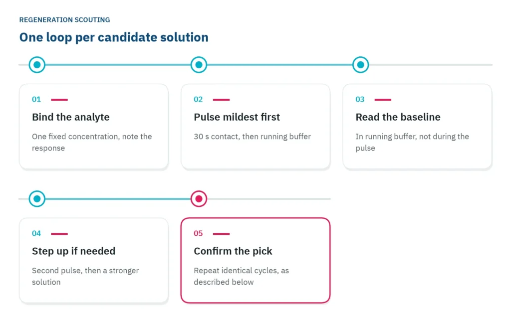
Biosensor surface regeneration is the step that lets one sensor surface serve many samples. In a label-free binding assay, the ligand sits on the sensor surface and the analyte flows over it. One cycle records binding, then dissociation in buffer, then a regeneration step that removes the analyte still bound, so the next cycle starts from clean ligand [1]. Most reuse problems start in that last step.
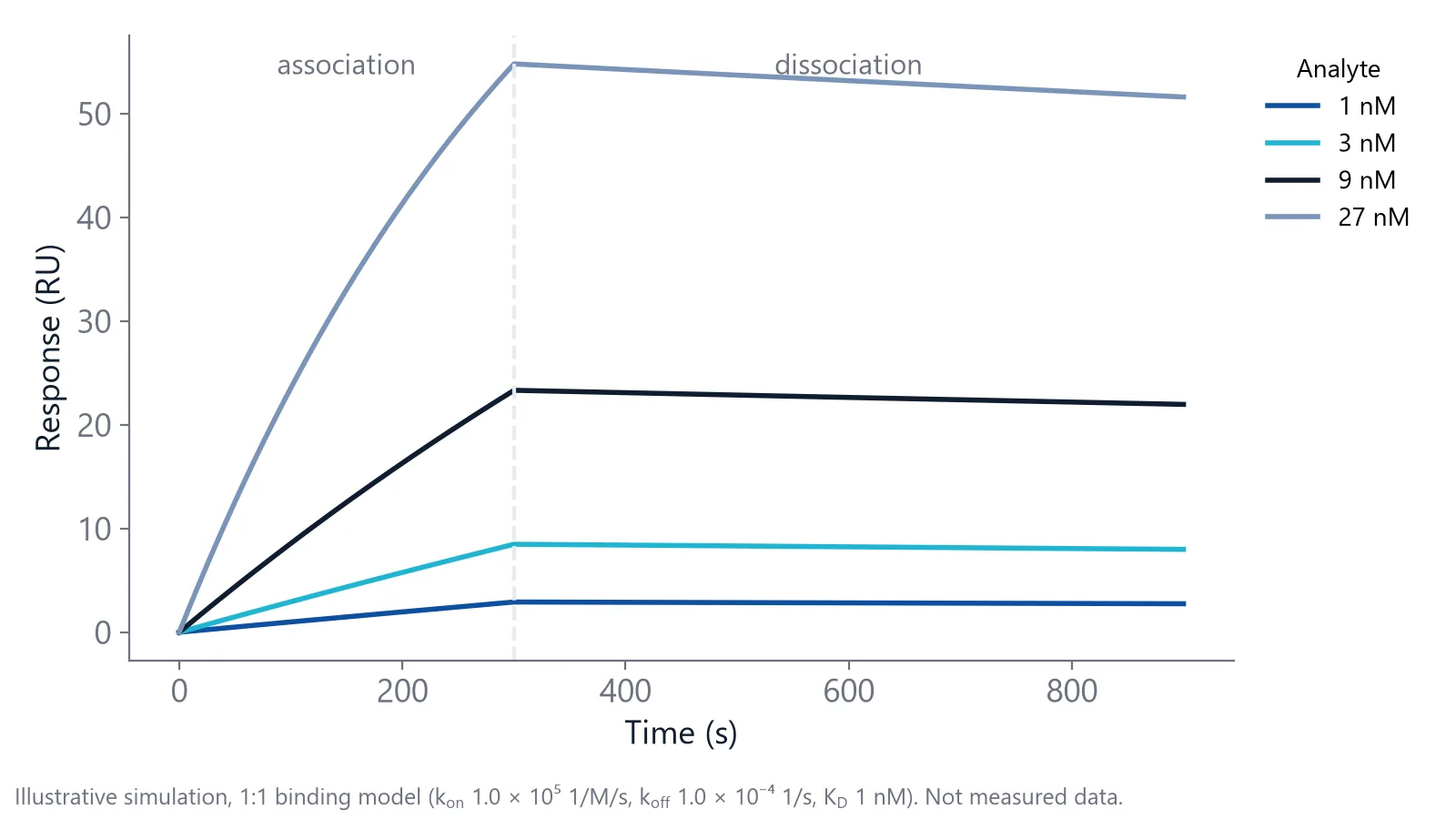
Buffer alone often cannot clear the surface. For a tight binder, a koff of 10-4 1/s means about 0.01% of the complexes fall apart every second. After 10 minutes of buffer, about 94% of the analyte is still there.
The regeneration solution has to break that complex without breaking the ligand. Miss in either direction and the data show it [1,2]:
- Too mild: analyte stays bound, so fewer sites are free for the next sample
- Too harsh: the ligand unfolds or comes off the surface
Both errors can pass for changing kinetics. Rmax, the maximum response, is proportional to the amount of active ligand, so a falling Rmax means fewer free active sites, from carryover or a damaged ligand, not a changing interaction [3].
Capture or titrate when the ligand cannot take it
Some complexes only let go under conditions that also destroy the ligand. Then change the format instead of the solution.
On a capture surface, a capture molecule such as Protein A, an anti-tag antibody or tris-NTA for His-tagged proteins holds the ligand. Regeneration removes ligand and analyte together, and fresh ligand goes on at the start of each cycle [4]. The solution now only has to spare the capture layer. This is the usual route for antibody affinity and kinetics work, where antibodies are held by their Fc region.
In single-cycle kinetics, you inject increasing concentrations one after another with short dissociations in between, and regenerate once at the end of the series. You still need blank cycles for referencing and drift [3].
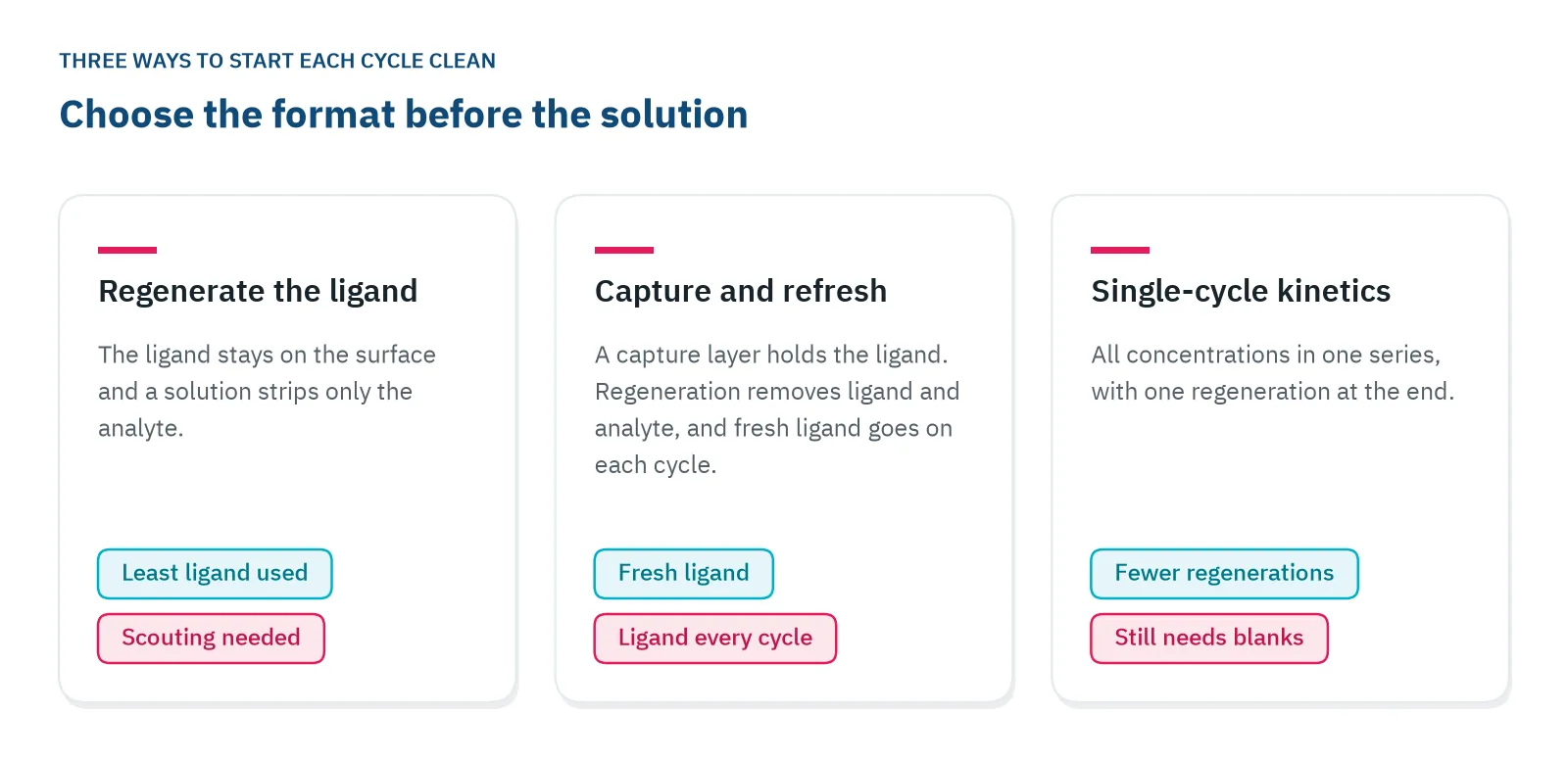
A fresh surface per measurement is the last resort. It costs chips and time, but it removes carryover completely.
Which regeneration solution should you try first?
Match the solution to the force that holds the complex together, and start at the mild end. Acid, base and high salt are the usual first choices, and two of them can be combined when one alone falls short. Denaturing agents are for very stable ligands only, at mild concentrations; treat organic solvents with the same care [2]. Typical starting values are in the table [1,2].
| Class | Typical solutions | Breaks mainly | Watch out |
|---|---|---|---|
| Low pH | 10 mM glycine-HCl, pH 3 down to 1.5; 10 to 100 mM HCl | Charge and hydrogen-bond contacts | Acid-sensitive ligands |
| High pH | 10 mM glycine-NaOH; up to 100 mM NaOH | Charge contacts, from the other side | Base-sensitive ligands |
| High salt | 1 to 5 M NaCl; 3 M MgCl2 | Electrostatic contacts | Large bulk signal during the pulse |
| Chelator | Ethylenediaminetetraacetic acid (EDTA) | Metal-dependent binding | Also strips metal from chelator surfaces |
| Detergent or organic | About 0.5% sodium dodecyl sulfate (SDS); about 50% ethylene glycol | Hydrophobic contacts | Stable ligands only |
Glycine-HCl at pH 2.5 and 3 M MgCl2 are the classic first tries, and one handbook suggests starting antibody and antigen pairs with the MgCl2 [1].
If no single solution works, mix them. In a screen of 13 antibody and antigen pairs, cocktails built from six stock solutions removed more than 90% of bound analyte in one 30 s pulse for 9 pairs. For 5 pairs they cleaned the surface fully under gentler conditions than those used before, and EDTA proved a useful additive to cocktails rich in chaotropic agents and salt [5].
Scout from mild to harsh
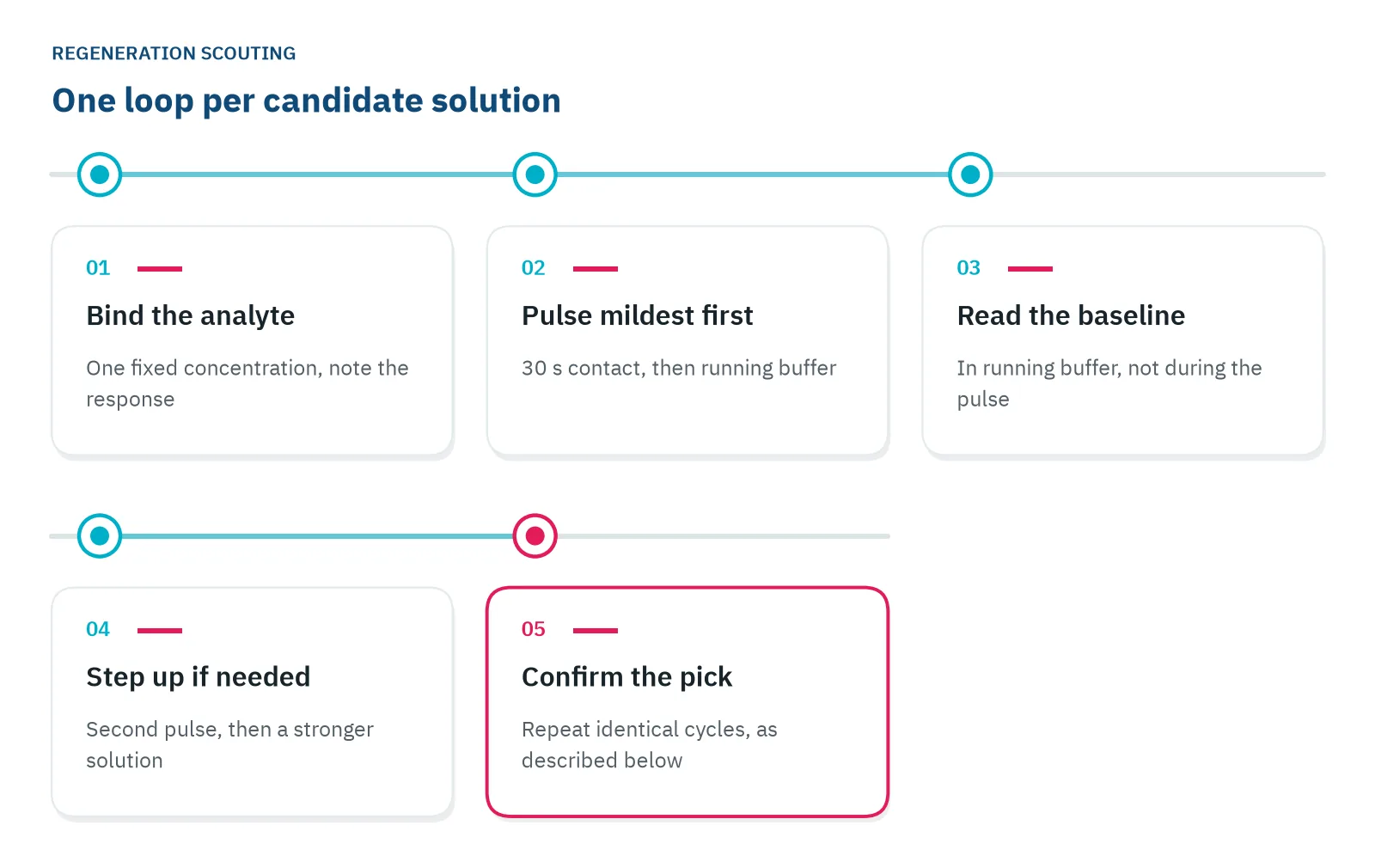
To regenerate the sensor surface without guessing, scout on a surface you can spare, with one analyte concentration that gives a clear response. Keep contact short: 30 s to 1 min is often enough, and two short pulses with buffer in between can be gentler on the ligand than one long pulse [1,2].
- Start with 10 mM glycine-HCl at pH 3, or the MgCl2 above for antibody pairs
- Lower the pH to 2.5, 2.0 and then 1.5
- Repeat the pulse once with buffer in between
- Move to a mixed solution
Ignore the signal during the pulse. Regeneration solutions differ in refractive index and can make the surface layer swell or shrink, so read the baseline in running buffer afterwards [1].
Stop at the mildest condition that returns the baseline to its starting level, then confirm it over repeated cycles, below.
How do you know surface regeneration on a biosensor worked?
Run the same sample through five to ten binding and regeneration cycles and plot two numbers per cycle: the baseline after regeneration, and the response to the sample. A rising baseline means analyte is left behind. A falling response means fewer free active sites, most often a ligand losing activity [1,2].
Both trends can show up together. On a nitrilotriacetic acid (NTA) surface, where regeneration removed His6-tagged proteins so fresh ones could be captured, the first recipe left the baseline climbing and the capture signal falling, a sign of protein building up. Adding a 3 min wash in 0.5 M NaOH improved removal, and the remaining loss over ten cycles depended on the protein [6].
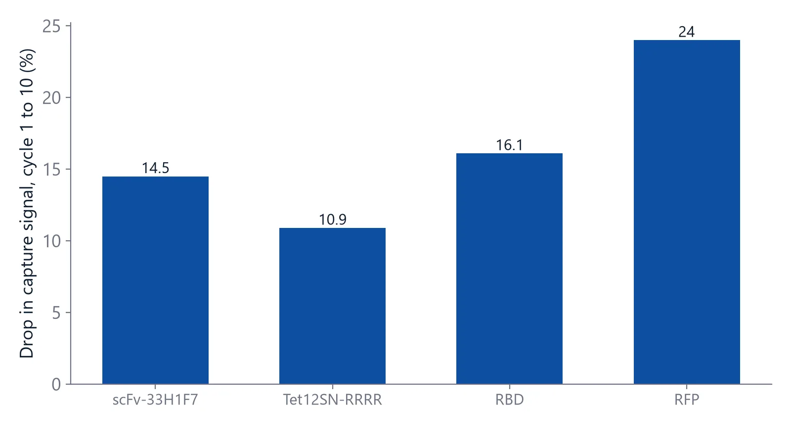
How much loss you can accept depends on the question. Concentration measurements read the response against a standard curve, so they need more complete regeneration than kinetic work [7]. For kinetics, randomize the concentration order: a series run from low to high can hide regeneration and drift problems [3].
Over a longer project, keep a control chart. In a long-term kinetic study of one antigen and antibody pair, regeneration conditions were among the main sources of variation, next to surface age and cycle count, which reached about 250 to 600 cycles per chip [8]. Regenerable sensor chips for inQuiQ are built for this kind of reuse, and the chip reusability and stability application note follows reuse over repeated analyses [9].
Find one regeneration that suits several ligands
When several ligands share one flow path or one array, they all get the same regeneration solution. Scout each ligand on its own first, then group the ligands that tolerate the same solution, one that is neither too mild nor too harsh for any of them [1]. If one ligand needs a much harsher step than the rest, give it its own run or move it to a capture format.
This matters for serial flow. In the standard setup on the inQuiQ benchtop biosensor, one sample injection flows through all four channels in turn, each with its own ligand and one serving as the reference, and the surface is regenerated once after that injection. To regenerate the sensor surface cleanly, put ligands on one chip that share a regeneration solution, and include the reference channel in your scouting, because it gets the same pulse.
Questions researchers ask
How many cycles can one sensor surface take?
Lifetime depends on the ligand stability and the regeneration conditions, and is typically tens to hundreds of cycles. A stable antibody can outlast a fragile enzyme. Track a control sample as in the control chart above and retire the surface when it falls below your limit.
Can I regenerate a streptavidin and biotin surface?
Conditions strong enough to break the streptavidin and biotin bond usually damage streptavidin too. Treat the biotinylated ligand as permanent and regenerate only the analyte off it. If you need fresh ligand for each measurement, choose a reversible capture instead, such as Protein A for antibodies or tris-NTA for His-tagged proteins.
Should I condition a new surface before the first cycle?
Yes. A fresh surface often changes most during its first contact with the regeneration solution. Run two or three regeneration pulses before the first real binding cycle, then a few buffer cycles, and leave them out of the analysis. Cycle one then behaves like the cycles that follow it.
Why does the baseline keep moving after a regeneration pulse?
Harsh solutions can leave the surface layer swollen or charged for a while. Add a buffer wash or a stabilization period before the next injection and check that the baseline is flat before injecting. Otherwise the drift ends up inside your association phase.
Do small molecule assays need regeneration?
Often they do not. Many small molecules and fragments dissociate within seconds to minutes, so the surface clears in buffer on its own. Check that the signal returns to baseline after each injection. If it does, skip regeneration and spare the ligand. Add a mild wash only for compounds that stick.
References
- Schasfoort RBM. Introduction to Surface Plasmon Resonance. Handbook of Surface Plasmon Resonance. 2017. doi:10.1039/9781788010283-00001
- Real-Time Analysis of Biomolecular Interactions. Springer Japan. 2000. doi:10.1007/978-4-431-66970-8
- Marquart JA. SPRpages – Getting a Feeling for the Curves. Handbook of Surface Plasmon Resonance. 2017. doi:10.1039/9781788010283-00106
- Gedig ET. Surface Chemistry in SPR Technology. Handbook of Surface Plasmon Resonance. 2017. doi:10.1039/9781788010283-00171
- Andersson K, Hämäläinen M, Malmqvist M. Identification and optimization of regeneration conditions for affinity-based biosensor assays. A multivariate cocktail approach. Anal Chem. 1999. doi:10.1021/ac981271j
- Qu JH, Leirs K, Escudero R et al.. Novel Regeneration Approach for Creating Reusable FO-SPR Probes with NTA Surface Chemistry. Nanomaterials (Basel). 2021. doi:10.3390/nano11010186
- Apiyo DO. Biolayer Interferometry (Octet) for Label-free Biomolecular Interaction Sensing. Handbook of Surface Plasmon Resonance. 2017. doi:10.1039/9781788010283-00356
- Steinicke F, Oltmann-Norden I, Wätzig H. Long term kinetic measurements revealing precision and general performance of surface plasmon resonance biosensors. Anal Biochem. 2017. doi:10.1016/j.ab.2017.05.009
- Delta Life Science. Chip Reusability and Stability. Application note. Link
Keep reading
 Baseline drift in a biosensor: why it happens and how to stop itBaseline drift is often blamed on the chip. A short buffer-only test shows whether the surface, the buffer or the instrument is the real cause.Assay setup
 His tag capture assay: what to check when capture levels driftHis-tag capture is fast and reversible, but the ligand keeps leaving the surface. How to measure that drift, keep it apart from the analyte interaction, and reduce it.Assay setup
 Bulk shift or binding? How to match buffers and read the jumpA square jump at the start of an injection is usually buffer, not binding. How to recognize a bulk shift, why the reference channel does not fully remove it, and how to match buffers.