
On this page
Questions about your own assay?
Ask a specialistIn a His tag capture assay, the captured ligand keeps leaving the surface, and that loss can look like analyte dissociation. Mono-NTA binds His6 only at micromolar affinity, so drift is common. Measure capture decay with blank cycles, keep high chelator and imidazole levels out of running buffer and samples, and stabilize capture with lower density, longer tags or tris-NTA.
Why capture levels drift in a His tag capture assay
A His tag capture assay holds the ligand on the sensor through its histidine tag instead of a covalent bond. The surface carries nickel-loaded nitrilotriacetic acid (Ni-NTA) or an anti-His antibody. Capture is reversible, so you can strip the surface and load fresh ligand for every cycle [1,2].
The price is stability. A single His tag binds Ni-NTA with a KD of about 1 µM at neutral pH [2]. Other work puts mono-NTA affinity for His6 near 10 µM [3]. At these affinities the ligand keeps leaving the surface. The result is a baseline that drifts down and a ligand density that falls during the run [1].
The surface works at all because released ligand often rebinds to free NTA groups nearby. Even so, most proteins with one His tag dissociate very fast, while two tags are usually enough for stable binding [2]. Captured ligand also reacts to pH, ionic strength and chelating additives [1].
How do you tell capture drift from analyte dissociation?
Run blank cycles. Capture the ligand exactly as in a sample cycle, then inject running buffer instead of analyte. The signal loss you see is capture decay alone. Subtract these blanks from the analyte cycles, and compare the capture-only decay rate with the dissociation you want to measure. If they are similar in size, fix the capture first.
On a capture surface, the dissociation phase mixes two processes: analyte leaving the ligand, and ligand (with any analyte still bound) leaving the surface [5]. Without correction, the second process makes koff look faster and can bend the curves away from a 1:1 model. A reference surface helps minimize artifacts [4], and bulk refractive index shifts are a typical example. Blank cycles remove the drift that remains. Using both is often called double referencing [5]. Single-cycle kinetics also needs blank cycles to compensate for drift [5].
Three checks tell you whether subtraction will be enough:
- The capture level is close to the same in every cycle
- The blank decay is slow compared with the analyte signal you measure
- The referenced baseline is flat before each injection [5]
A simple example with made-up numbers shows the scale. You capture 400 RU of ligand, and the blank cycles lose 3 RU per minute. Over a 10 minute dissociation phase that is 30 RU. If your analyte gives a 30 RU signal and dissociates slowly, the capture decay is as large as what you are trying to measure.
When decay is large, small differences between cycles become errors in koff, so a more stable capture is the better fix.
Compare capture chemistries before you optimize
The table compares the common options for His-tagged ligands on surface plasmon resonance (SPR) and similar label-free biosensors.
| Chemistry | Affinity for the tag | Capture stability | Main trade-off |
|---|---|---|---|
| Mono-NTA (Ni²⁺) | About 1 to 10 µM for His6 [2,3] | Drift is common; rebinding helps | Hard for small or weak analytes [1] |
| Tris-NTA or poly-NTA | About 10 nM to subnanomolar [3,6] | Dissociation about 4 orders of magnitude slower than mono-NTA in solution [6] | Affinity depends on spacer design [3]; can raise non-specific binding and needs a special surface [7] |
| Anti-His antibody | Depends on the antibody | Varies from ligand to ligand; fewer regeneration cycles than NTA [7] | Large IgG takes up sensing volume and can add non-specific binding [1] |
| Capture, then covalent coupling | Permanent after coupling | No capture drift | Ligand cannot be stripped, so analyte regeneration must be optimized [1] |
Tris-NTA and poly-NTA surfaces regenerate under the same mild conditions as mono-NTA [1]. They need no change to the ligand, a practical upgrade when mono-NTA drifts [7].
Buffer and sample conditions that strip the tag
Ni-NTA capture depends on a metal complex, and many common additives compete with it. On NTA surfaces, keep divalent cations and significant amounts of chelating agents out of the running buffer [8]. Check these sources in particular:
- EDTA or EGTA carried over from protease inhibitor mixes or storage buffers
- Imidazole left in the protein after elution from a nickel column [1]
- Alkaline buffers, because NTA is unstable at high pH [8]
- Large differences in pH or salt between running buffer and sample [1]
Low levels can help: one protocol adds 50 µM EDTA and 5 mM imidazole to the running buffer to reduce non-specific binding [8]. Desalt the ligand into running buffer before capture, and prepare the analyte in the same buffer. Charge the NTA with nickel at the start of each cycle (for example with 500 µM NiCl₂). Strip it with a high EDTA concentration at the end, which removes nickel and ligand together [8].
For the running buffer itself, HEPES gave the best results in one handbook protocol, while PBS and Tris also worked [1]. Early drift often comes from a surface that has not equilibrated, so run a few buffer injections before the first analyte [5].
How can you make His-tag capture more stable?
Lower the capture level, use a longer or double His tag, or move to a multivalent chelator such as tris-NTA. Or capture it and then couple it covalently. Capture fresh ligand in every cycle. Single-cycle kinetics needs fewer capture and regeneration steps, but one capture must then last the whole series, so use it only if drift is low.
Lower the ligand density
A lower capture level leaves more free NTA groups, which raises the chance that a released ligand rebinds [1]. Set the level with ligand concentration and contact time [8]. A low density also limits mass transport effects and analyte rebinding [2].
Change the tag
A decahistidine (His10) tag was captured more stably than His6, and a double tag did better still. In one test of a His6 and a His10 tag joined by a spacer, average drift over two days of continuous flow was about 2 RU per minute. The handbook authors judge that low enough to correct for, even in small molecule work [1]. Whether your drift is acceptable depends on your analyte signal, so compare it with your own blank cycles.
Use a multivalent chelator
Only two of the six histidines in a His6 tag coordinate one Ni-NTA group. Tris-NTA engages the others and binds about three orders of magnitude more tightly [1].
Fix the ligand, or refresh it
Capture followed by amine coupling removes capture drift. The ligand can then no longer be stripped, so you need a regeneration that removes analyte and spares ligand [1]. The opposite route is to capture fresh ligand every cycle, which also removes ligand damage from regeneration as a source of error [2]. Single-cycle kinetics needs only one regeneration per concentration series, but one capture must last the whole series, and blank cycles are still needed to correct drift [5].
A check routine before you fit
Run this sequence during assay development, before kinetic data.
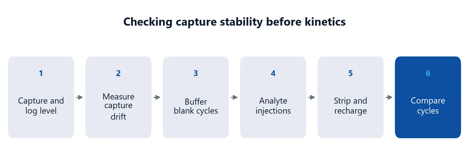
- Capture the ligand and record the capture level
- Wait in running buffer and measure capture drift
- Run buffer blanks with the same capture step
- Inject the analyte series
- Strip with EDTA and recharge with nickel
- Compare capture levels and blank decay across cycles
If capture levels fall from cycle to cycle, suspect loss of surface capacity or chelators in the samples. If the blank decay is large, change the chemistry or the tag before you fit. Curves of low quality give wrong rate constants however well you fit them, so optimize the surface first [5].
Checking capture stability on inQuiQ
Capture assays depend on telling ligand loss apart from instrument drift. On the inQuiQ benchtop biosensor, baseline drift is typically below 0.3 RU/min, and NTA capture is among the supported chemistries for His-tagged proteins. With in-flow immobilization, each of the 4 channels carries its own ligand. A His6 and a His10 construct can then be captured side by side, with a channel without ligand as the reference.
Delta's regenerable sensor chips can be reused across runs, as the chip reusability and stability application note shows [9]. That note concerns chips in general, not His-tag capture.
Questions researchers ask
Does imidazole in my protein sample affect His-tag capture?
Yes. Imidazole competes with the histidine tag for the nickel, so the high imidazole used for elution from a nickel column can lower capture levels and speed up ligand loss. Desalt or dialyze the protein into running buffer before capture, and use the same buffer for the analyte to avoid mismatches.
Can I measure small molecule binding on a Ni-NTA capture surface?
It is difficult on mono-NTA. Small or weak analytes give small signals, and baseline drift plus falling ligand density can hide them. If you need capture, use a multivalent chelator or a longer tag and check that the remaining drift is low enough to correct. Otherwise consider covalent or capture-then-couple immobilization.
How many times can I regenerate an NTA surface?
On a good quality NTA surface, around 100 strip cycles with EDTA have been reported without a clear loss of capacity. Because the ligand leaves with the nickel, one surface can host many different His-tagged proteins. Track the capture level per cycle so you notice when capacity starts to fall.
Why does my capture level drop between cycles even with fresh ligand?
Common causes are incomplete nickel recharge, chelators or imidazole in the samples, and loss of surface capacity. Traces of other metal ions in the flow system can compete with nickel, so clean the system with EDTA the day before and then keep running buffer flowing.
When is anti-His antibody capture a better choice than Ni-NTA?
Consider it when your buffer or sample must contain chelators or metal ions, because antibody capture does not rely on nickel. Stability still varies from ligand to ligand, and the large antibody can add background and takes up sensing volume. Test both with your own ligand.
References
- Gedig ET. Surface Chemistry in SPR Technology. Handbook of Surface Plasmon Resonance. 2017. doi:10.1039/9781788010283-00171
- Nieba L, Nieba-Axmann SE, Persson A et al.. BIACORE analysis of histidine-tagged proteins using a chelating NTA sensor chip. Anal Biochem. 1997. doi:10.1006/abio.1997.2326
- Huang Z, Hwang P, Watson DS et al.. Tris-nitrilotriacetic acids of subnanomolar affinity toward hexahistidine tagged molecules. Bioconjug Chem. 2009. doi:10.1021/bc900309n
- Myszka DG. Improving biosensor analysis. J Mol Recognit. 1999. doi:10.1002/(SICI)1099-1352(199909/10)12:5<279::AID-JMR473>3.0.CO;2-3
- Marquart JA. SPRpages – Getting a Feeling for the Curves. Handbook of Surface Plasmon Resonance. 2017. doi:10.1039/9781788010283-00106
- Lata S, Reichel A, Brock R et al.. High-affinity adaptors for switchable recognition of histidine-tagged proteins. J Am Chem Soc. 2005. doi:10.1021/ja050690c
- Questions and Answers. Handbook of Surface Plasmon Resonance. 2017. doi:10.1039/9781788010283-00479
- Real-Time Analysis of Biomolecular Interactions. Springer Japan. 2000. doi:10.1007/978-4-431-66970-8
- Delta Life Science. Chip Reusability and Stability. Application note. Link
Keep reading
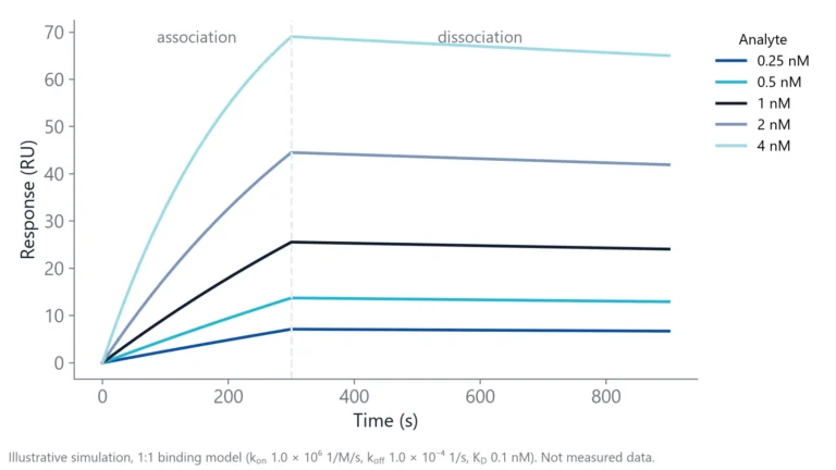 Baseline drift in a biosensor: why it happens and how to stop itBaseline drift is often blamed on the chip. A short buffer-only test shows whether the surface, the buffer or the instrument is the real cause.Assay setup
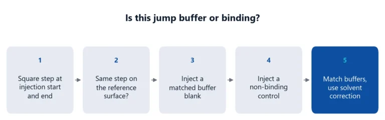 Bulk shift or binding? How to match buffers and read the jumpA square jump at the start of an injection is usually buffer, not binding. How to recognize a bulk shift, why the reference channel does not fully remove it, and how to match buffers.Applications
 Matrix effects in binding assays: how serum changes your signalSerum can raise, lower or blur the signal of a binding assay. Learn where matrix effects come from, how to test for them, and how to reduce them in ELISA and label-free assays.